Deposition Date
2023-03-03
Release Date
2023-08-30
Last Version Date
2024-11-13
Entry Detail
PDB ID:
8CQ4
Keywords:
Title:
Bifunctional cyclohexadienyl dehydratase/chorismate mutase from Janthinobacterium sp. HH01
Biological Source:
Source Organism(s):
Janthinobacterium sp. HH01 (Taxon ID: 1198452)
Expression System(s):
Method Details:
Experimental Method:
Resolution:
1.65 Å
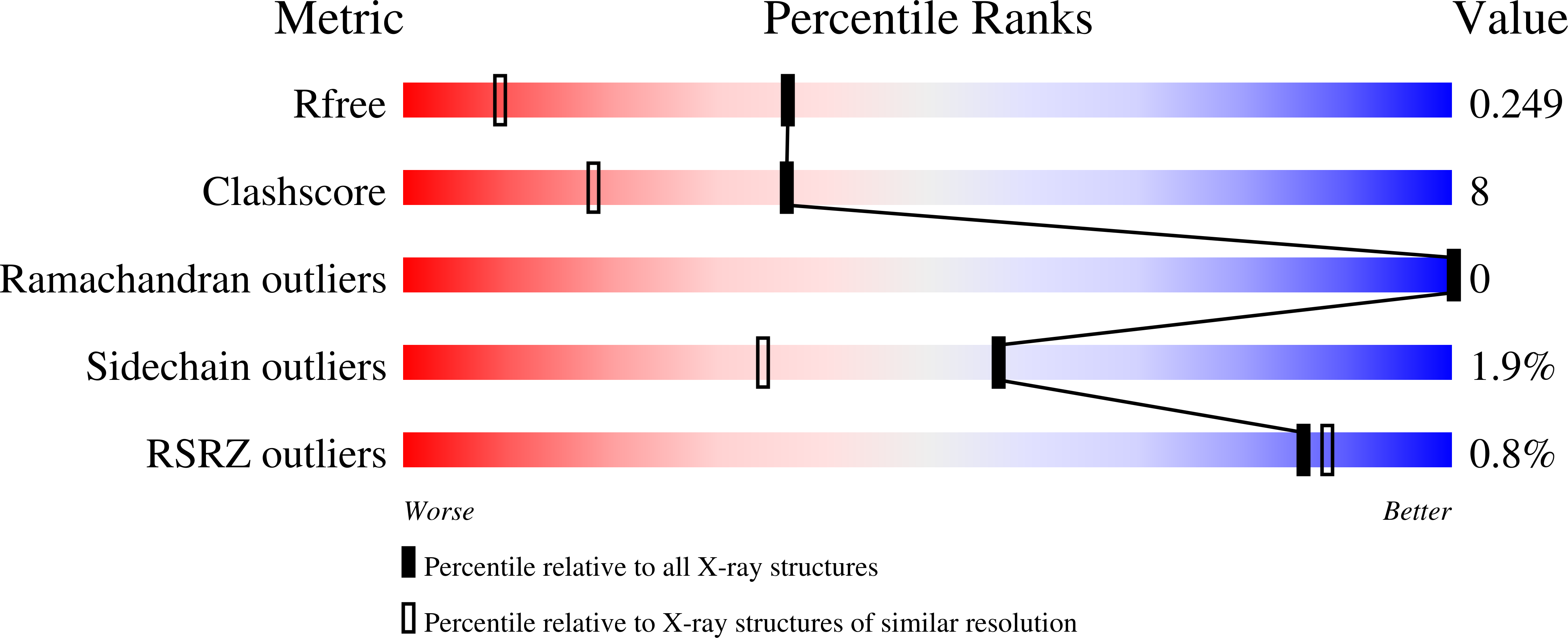
R-Value Free:
0.24
R-Value Work:
0.19
Space Group:
P 21 21 21