8CPQ image
Deposition Date 2023-03-03
Release Date 2024-03-13
Last Version Date 2025-09-24
Entry Detail
PDB ID:
8CPQ
Keywords:
Title:
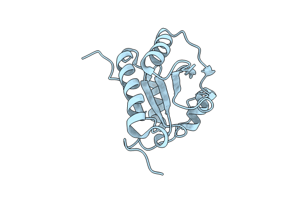
Crystal structure of human protein disulfide isomerase PDIA6 domain b
Biological Source:
Source Organism:
Homo sapiens (Taxon ID: 9606)
Method Details:
Experimental Method:
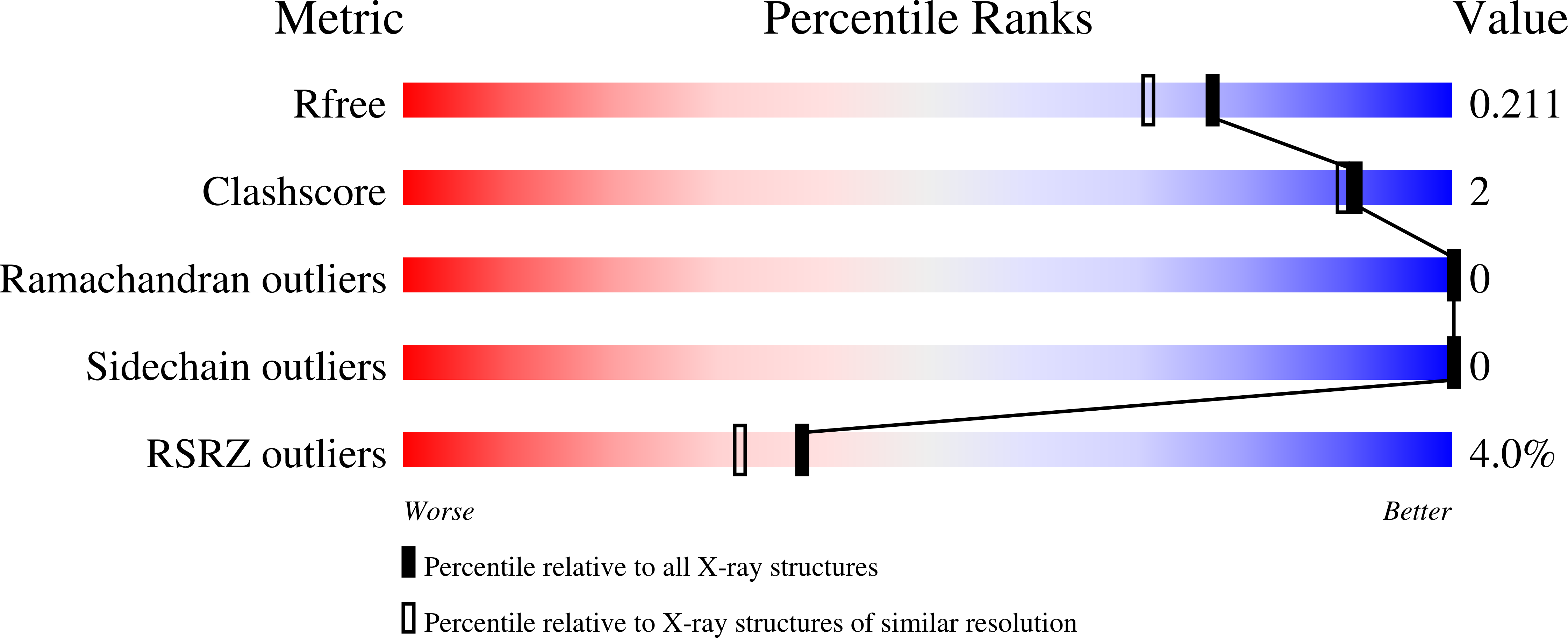
Resolution:
1.80 Å
R-Value Free:
0.21
R-Value Work:
0.17
R-Value Observed:
0.17
Space Group:
C 2 2 21
Macromolecular Entities
Structures with similar UniProt ID
Protein Blast
Polymer Type:polypeptide(L)
Molecule:Protein disulfide-isomerase A6
Gene (Uniprot):PDIA6
Chain IDs:A
Chain Length:174
Number of Molecules:1
Biological Source:Homo sapiens
Primary Citation
A multichaperone condensate enhances protein folding in the endoplasmic reticulum.
Nat.Cell Biol. 27 1422 1430 (2025)
PMID: 40789936 DOI: 10.1038/s41556-025-01730-w

Abstact

Protein folding in the endoplasmic reticulum (ER) relies on a network of molecular chaperones that facilitates the folding and maturation of client proteins. How the ER chaperones organize in a supramolecular manner to exert their cooperativity has, however, remained unclear. Here we report the discovery of a multichaperone condensate in the ER lumen, which is formed around the chaperone PDIA6 during protein folding homeostasis. The condensates form in a Ca2+-dependent manner and we resolve the underlying mechanism at the atomic and cellular levels. The PDIA6 condensates recruit further chaperones-Hsp70 BiP, J-domain protein ERdj3, disulfide isomerase PDIA1 and Hsp90 Grp94-which constitute some of the essential components of the early folding machinery. The chaperone condensates enhance folding of proteins, such as proinsulin, and prevent protein misfolding in the ER lumen. The PDIA6-scaffolded chaperone condensates hence provide the functional basis for spatial and temporal coordination of the dynamic ER chaperone network.

Legend

Protein

Chemical

Disease

Primary Citation of related structures