8CPE image
Deposition Date 2023-03-02
Release Date 2023-08-16
Last Version Date 2024-11-06
Entry Detail
PDB ID:
8CPE
Keywords:
Title:
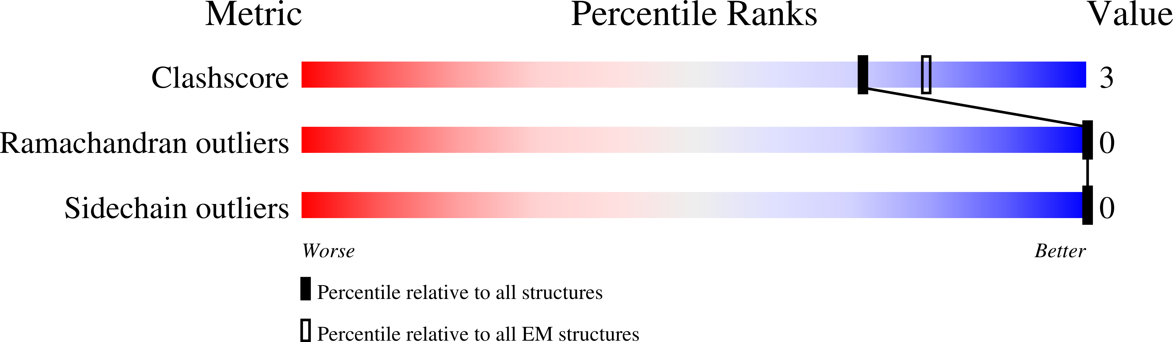
CryoEM structure of AL55 amyloid fibrils extracted from the kidney of an AL amyloidosis patient.
Biological Source:
Source Organism:
Homo sapiens (Taxon ID: 9606)
Method Details:
Experimental Method:
Resolution:
4.00 Å
Aggregation State:
FILAMENT
Reconstruction Method:
HELICAL
Macromolecular Entities
Polymer Type:polypeptide(L)
Molecule:Immunoglobulin lambda light chain
Mutations:No
Chain IDs:A (auth: C), B (auth: A), C (auth: B), D, E
Chain Length:217
Number of Molecules:5
Biological Source:Homo sapiens
Ligand Molecules
Primary Citation
The Cryo-EM STRUCTURE of Renal Amyloid Fibril Suggests Structurally Homogeneous Multiorgan Aggregation in AL Amyloidosis.
J.Mol.Biol. 435 168215 168215 (2023)
PMID: 37516426 DOI: 10.1016/j.jmb.2023.168215

Abstact

Immunoglobulin light chain amyloidosis (AL) is caused by the aberrant production of amyloidogenic light chains (LC) that accumulate as amyloid deposits in vital organs. Distinct LC sequences in each patient yield distinct amyloid structures. However different tissue microenvironments may also cause identical protein precursors to adopt distinct amyloid structures. To address the impact of the tissue environment on the structural polymorphism of amyloids, we extracted fibrils from the kidney of an AL patient (AL55) whose cardiac amyloid structure was previously determined by our group. Here we show that the 4.0 Å resolution cryo-EM structure of the renal fibril is virtually identical to that reported for the cardiac fibril. These results provide the first structural evidence that LC amyloids independently deposited in different organs of the same AL patient share a common fold.

Legend

Protein

Chemical

Disease

Primary Citation of related structures