8CP0 image
Entry Detail
PDB ID:
8CP0
Keywords:
Title:
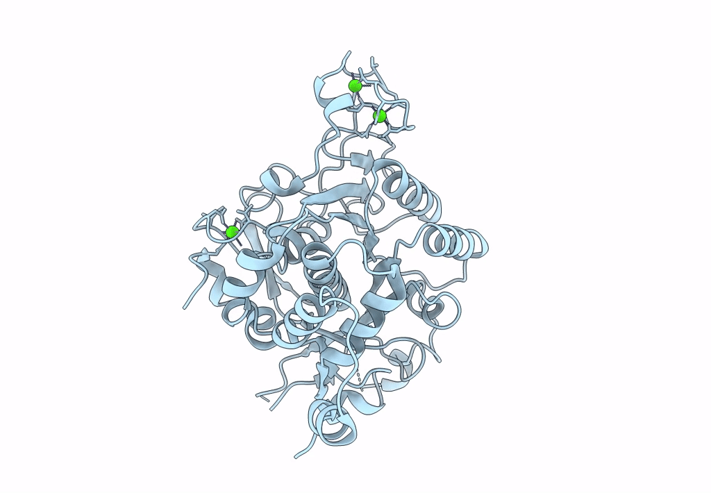
Structure of the catalytic domain of P. vivax Sub1 (trigonal crystal form)
Biological Source:
Source Organism:
PDB Version:
Deposition Date:
2023-03-01
Release Date:
2023-07-19
Method Details:
Experimental Method:
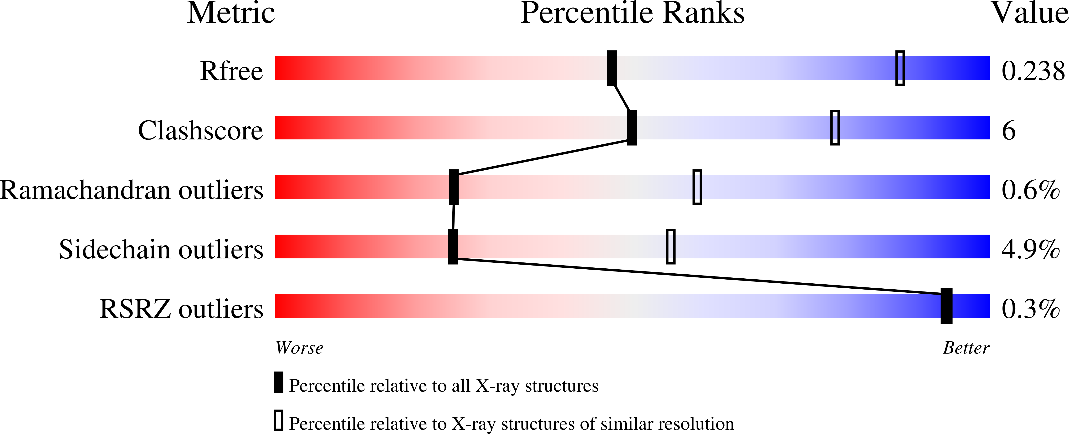
Resolution:
3.25 Å
R-Value Free:
0.24
R-Value Work:
0.19
R-Value Observed:
0.19
Space Group:
P 32 2 1
Macromolecular Entities
Polymer Type:polypeptide(L)
Description:subtilisin
Chain IDs:A
Chain Length:393
Number of Molecules:1
Biological Source:Plasmodium vivax
Ligand Molecules
Primary Citation
3D structures of the Plasmodium vivax subtilisin-like drug target SUB1 reveal conformational changes to accommodate a substrate-derived alpha-ketoamide inhibitor.
Acta Crystallogr D Struct Biol 79 721 734 (2023)
PMID: 37428845 DOI: 10.1107/S2059798323004710

Abstact

The constant selection and propagation of multi-resistant Plasmodium sp. parasites require the identification of new antimalarial candidates involved in as-yet untargeted metabolic pathways. Subtilisin-like protease 1 (SUB1) belongs to a new generation of drug targets because it plays a crucial role during egress of the parasite from infected host cells at different stages of its life cycle. SUB1 is characterized by an unusual pro-region that tightly interacts with its cognate catalytic domain, thus precluding 3D structural analysis of enzyme-inhibitor complexes. In the present study, to overcome this limitation, stringent ionic conditions and controlled proteolysis of recombinant full-length P. vivax SUB1 were used to obtain crystals of an active and stable catalytic domain (PvS1Cat) without a pro-region. High-resolution 3D structures of PvS1Cat, alone and in complex with an α-ketoamide substrate-derived inhibitor (MAM-117), showed that, as expected, the catalytic serine of SUB1 formed a covalent bond with the α-keto group of the inhibitor. A network of hydrogen bonds and hydrophobic interactions stabilized the complex, including at the P1' and P2' positions of the inhibitor, although P' residues are usually less important in defining the substrate specificity of subtilisins. Moreover, when associated with a substrate-derived peptidomimetic inhibitor, the catalytic groove of SUB1 underwent significant structural changes, particularly in its S4 pocket. These findings pave the way for future strategies for the design of optimized SUB1-specific inhibitors that may define a novel class of antimalarial candidates.

Legend

Protein

Chemical

Disease

Primary Citation of related structures