8CM9 image
Entry Detail
PDB ID:
8CM9
Keywords:
Title:
Structure of human O-GlcNAc transferase in complex with UDP and tP11
Biological Source:
PDB Version:
Deposition Date:
2023-02-18
Release Date:
2023-10-04
Method Details:
Experimental Method:
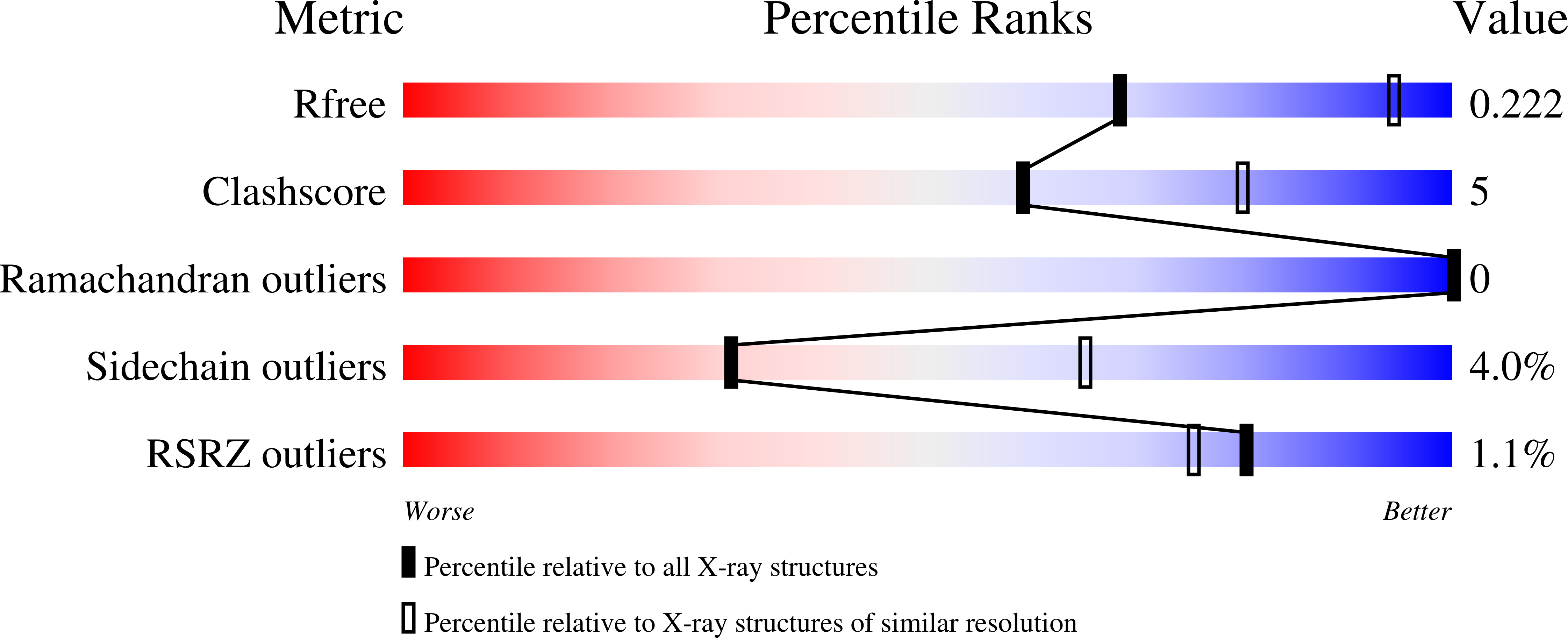
Resolution:
2.80 Å
R-Value Free:
0.22
R-Value Work:
0.19
Space Group:
P 3 2 1
Macromolecular Entities
Polymer Type:polypeptide(L)
Description:UDP-N-acetylglucosamine--peptide N-acetylglucosaminyltransferase 110 kDa subunit
Chain IDs:A, B, C, D
Chain Length:723
Number of Molecules:4
Biological Source:Homo sapiens
Polymer Type:polypeptide(L)
Description:PHE-MET-PRO-LYS-TYR-SER-ILE
Chain IDs:E, F, G, H
Chain Length:7
Number of Molecules:4
Biological Source:synthetic construct
Ligand Molecules
Primary Citation
Phage display uncovers a sequence motif that drives polypeptide binding to a conserved regulatory exosite of O-GlcNAc transferase.
Proc.Natl.Acad.Sci.USA 120 e2303690120 e2303690120 (2023)
PMID: 37819980 DOI: 10.1073/pnas.2303690120

Abstact

The modification of nucleocytoplasmic proteins by O-linked N-acetylglucosamine (O-GlcNAc) is an important regulator of cell physiology. O-GlcNAc is installed on over a thousand proteins by just one enzyme, O-GlcNAc transferase (OGT). How OGT is regulated is therefore a topic of interest. To gain insight into these questions, we used OGT to perform phage display selection from an unbiased library of ~109 peptides of 15 amino acids in length. Following rounds of selection and deep mutational panning, we identified a high-fidelity peptide consensus sequence, [Y/F]-x-P-x-Y-x-[I/M/F], that drives peptide binding to OGT. Peptides containing this sequence bind to OGT in the high nanomolar to low micromolar range and inhibit OGT in a noncompetitive manner with low micromolar potencies. X-ray structural analyses of OGT in complex with a peptide containing this motif surprisingly revealed binding to an exosite proximal to the active site of OGT. This structure defines the detailed molecular basis driving peptide binding and explains the need for specific residues within the sequence motif. Analysis of the human proteome revealed this motif within 52 nuclear and cytoplasmic proteins. Collectively, these data suggest a mode of regulation of OGT by which polypeptides can bind to this exosite to cause allosteric inhibition of OGT through steric occlusion of its active site. We expect that these insights will drive improved understanding of the regulation of OGT within cells and enable the development of new chemical tools to exert fine control over OGT activity.

Legend

Protein

Chemical

Disease

Primary Citation of related structures