8CBY image
Entry Detail
PDB ID:
8CBY
Keywords:
Title:
Crystal Structure of Anti-cortisol Fab in Complex with Cortisol
Biological Source:
Source Organism:
Host Organism:
PDB Version:
Deposition Date:
2023-01-26
Release Date:
2023-05-10
Method Details:
Experimental Method:
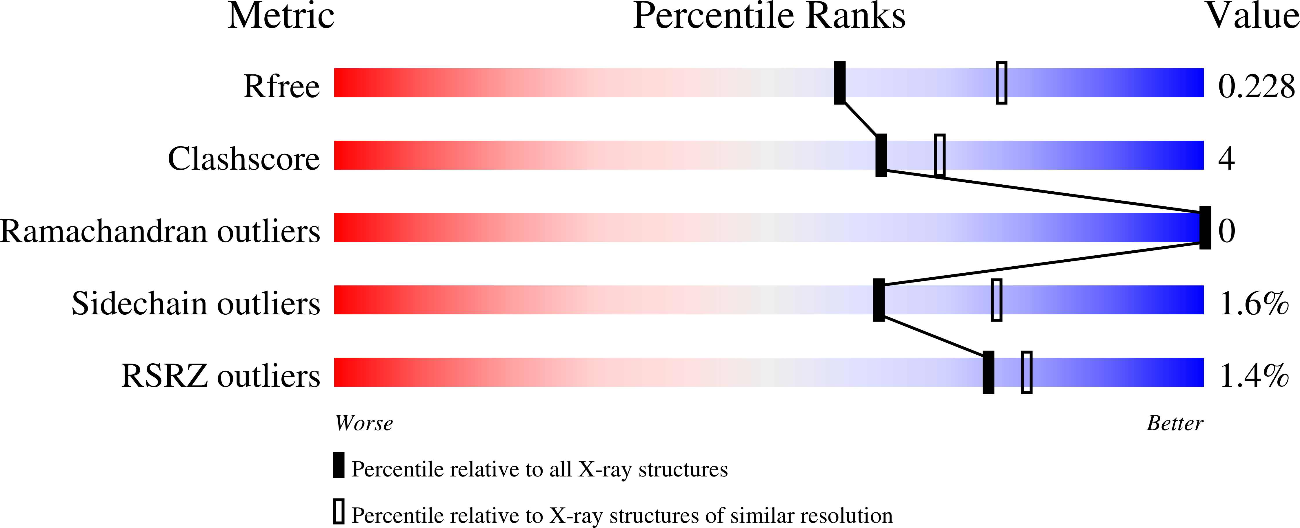
Resolution:
2.27 Å
R-Value Free:
0.23
R-Value Work:
0.20
R-Value Observed:
0.20
Space Group:
P 65 2 2
Macromolecular Entities
Polymer Type:polypeptide(L)
Description:anti-cortisol (17) Fab (heavy chain)
Chain IDs:B (auth: H)
Chain Length:230
Number of Molecules:1
Biological Source:Mus musculus
Polymer Type:polypeptide(L)
Description:anti-cortisol (17) Fab (light chain)
Chain IDs:A (auth: L)
Chain Length:211
Number of Molecules:1
Biological Source:Mus musculus
Ligand Molecules
Primary Citation
Structural insight to elucidate the binding specificity of the anti-cortisol Fab fragment with glucocorticoids.
J.Struct.Biol. 215 107966 107966 (2023)
PMID: 37100101 DOI: 10.1016/j.jsb.2023.107966

Abstact

Cortisol is a steroid hormone that is produced by the adrenal gland. It is a primary stress hormone that increases glucose levels in the blood stream. High concentrations of cortisol in the body can be used as a biomarker for acute and chronic stress and related mental and physiological disorders. Therefore, the accurate quantification of cortisol levels in body fluids is essential for clinical diagnosis. In this article, we describe the isolation of recombinant anti-cortisol antibodies with high affinity for cortisol and discover their cross-reactivity with other glucocorticoids. To describe the cortisol binding site and elucidate the structural basis for the binding specificity, the high-resolution crystal structures of the anti-cortisol (17) Fab fragment in the absence of glucocorticoid (2.00 Å) and the presence of cortisol (2.26 Å), corticosterone (1.86 Å), cortisone (1.85 Å) and prednisolone (2.00 Å) were determined. To our knowledge, this is the first determined crystal structure of a cortisol-specific antibody. The recognition of cortisol is driven by hydrophobic interactions and hydrogen bonding at the protein-ligand interface coupled with a conformational transition. Comparison of ligand-free and ligand-bound structures showed that the side chains of residues Tyr58-H and Arg56-H can undergo local conformational changes at the binding site, most likely prior to the binding event via a conformational selection mechanism. Compared to other anti-steroid antibody-antigen complexes, (17) Fab possesses a structurally unique steroid binding site, as the H3 loop from the CDR area has only a minor contribution, but framework residues have a prominent contribution to hapten binding.

Legend

Protein

Chemical

Disease

Primary Citation of related structures