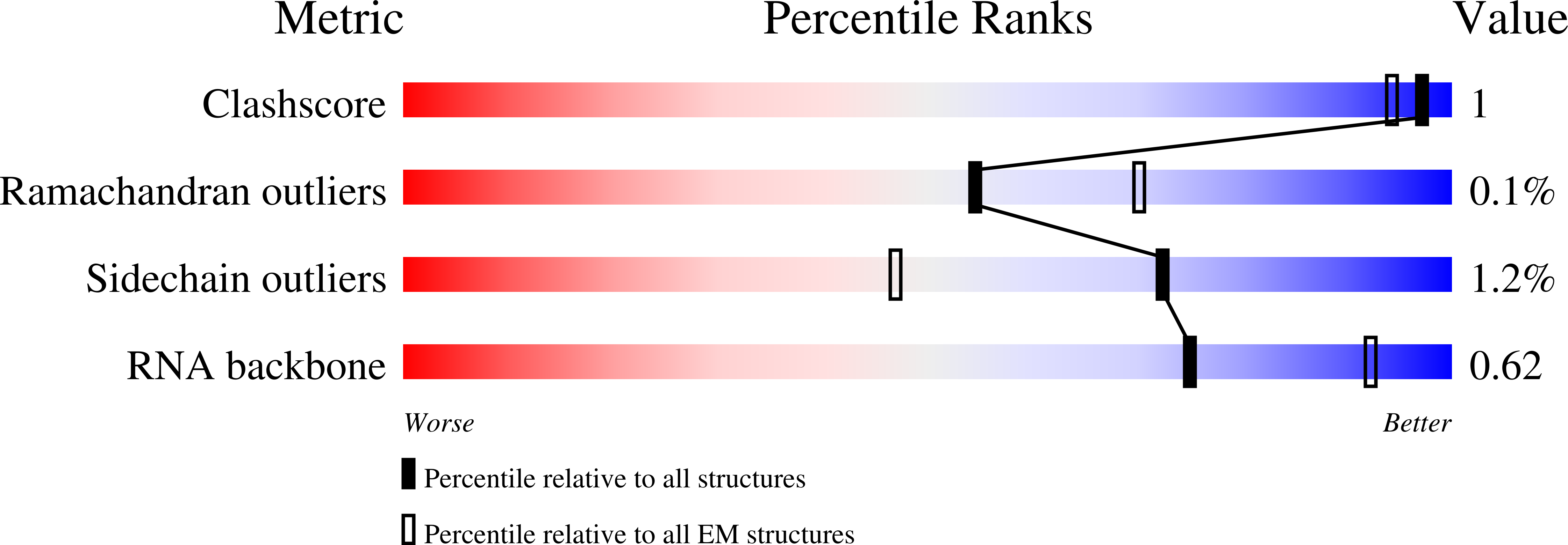
8CAI image
Deposition Date 2023-01-24
Release Date 2023-07-26
Last Version Date 2024-04-24
Entry Detail
PDB ID:
8CAI
Keywords:
Title:
Streptomycin and Hygromycin B bound to the 30S body
Biological Source:
Source Organism:
Method Details:
Experimental Method:
Resolution:
2.08 Å
Aggregation State:
PARTICLE
Reconstruction Method:
SINGLE PARTICLE
Macromolecular Entities
Polymer Type:polyribonucleotide
Molecule:16S rRNA
Chain IDs:A
Chain Length:1540
Number of Molecules:1
Biological Source:Escherichia coli BW25113
Polymer Type:polypeptide(L)
Molecule:30S ribosomal protein S2
Gene (Uniprot):rpsB
Chain IDs:B
Chain Length:241
Number of Molecules:1
Biological Source:Escherichia coli BW25113
Polymer Type:polypeptide(L)
Molecule:Small ribosomal subunit protein uS3
Gene (Uniprot):rpsC
Chain IDs:C
Chain Length:233
Number of Molecules:1
Biological Source:Escherichia coli BW25113
Polymer Type:polypeptide(L)
Molecule:Small ribosomal subunit protein uS4
Gene (Uniprot):rpsD
Chain IDs:D
Chain Length:206
Number of Molecules:1
Biological Source:Escherichia coli BW25113
Polymer Type:polypeptide(L)
Molecule:Small ribosomal subunit protein uS5
Gene (Uniprot):rpsE
Chain IDs:E
Chain Length:167
Number of Molecules:1
Biological Source:Escherichia coli BW25113
Polymer Type:polypeptide(L)
Molecule:Small ribosomal subunit protein bS6, non-modified isoform
Gene (Uniprot):rpsF
Chain IDs:F
Chain Length:131
Number of Molecules:1
Biological Source:Escherichia coli BW25113
Polymer Type:polypeptide(L)
Molecule:Small ribosomal subunit protein uS8
Gene (Uniprot):rpsH
Chain IDs:G (auth: H)
Chain Length:130
Number of Molecules:1
Biological Source:Escherichia coli BW25113
Polymer Type:polypeptide(L)
Molecule:Small ribosomal subunit protein uS11
Gene (Uniprot):rpsK
Chain IDs:H (auth: K)
Chain Length:129
Number of Molecules:1
Biological Source:Escherichia coli BW25113
Polymer Type:polypeptide(L)
Molecule:Small ribosomal subunit protein uS12
Gene (Uniprot):rpsL
Chain IDs:I (auth: L)
Chain Length:124
Number of Molecules:1
Biological Source:Escherichia coli BW25113
Polymer Type:polypeptide(L)
Molecule:Small ribosomal subunit protein uS15
Gene (Uniprot):rpsO
Chain IDs:J (auth: O)
Chain Length:89
Number of Molecules:1
Biological Source:Escherichia coli BW25113
Polymer Type:polypeptide(L)
Molecule:30S ribosomal protein S16
Gene (Uniprot):rpsP
Chain IDs:K (auth: P)
Chain Length:82
Number of Molecules:1
Biological Source:Escherichia coli BW25113
Polymer Type:polypeptide(L)
Molecule:Small ribosomal subunit protein uS17
Gene (Uniprot):rpsQ
Chain IDs:L (auth: Q)
Chain Length:84
Number of Molecules:1
Biological Source:Escherichia coli BW25113
Polymer Type:polypeptide(L)
Molecule:Small ribosomal subunit protein bS18
Gene (Uniprot):rpsR
Chain IDs:M (auth: R)
Chain Length:75
Number of Molecules:1
Biological Source:Escherichia coli BW25113
Polymer Type:polypeptide(L)
Molecule:30S ribosomal protein S20
Gene (Uniprot):rpsT
Chain IDs:N (auth: T)
Chain Length:87
Number of Molecules:1
Biological Source:Escherichia coli BW25113
Polymer Type:polypeptide(L)
Molecule:Small ribosomal subunit protein bS21
Gene (Uniprot):rpsU
Chain IDs:O (auth: U)
Chain Length:71
Number of Molecules:1
Biological Source:Escherichia coli BW25113
Modified Residue
Compound ID Chain ID Parent Comp ID Details 2D Image
D2T I ASP modified residue
Primary Citation
Structural conservation of antibiotic interaction with ribosomes.
Nat.Struct.Mol.Biol. 30 1380 1392 (2023)
PMID: 37550453 DOI: 10.1038/s41594-023-01047-y

Abstact

The ribosome is a major target for clinically used antibiotics, but multidrug resistant pathogenic bacteria are making our current arsenal of antimicrobials obsolete. Here we present cryo-electron-microscopy structures of 17 distinct compounds from six different antibiotic classes bound to the bacterial ribosome at resolutions ranging from 1.6 to 2.2 Å. The improved resolution enables a precise description of antibiotic-ribosome interactions, encompassing solvent networks that mediate multiple additional interactions between the drugs and their target. Our results reveal a high structural conservation in the binding mode between antibiotics with the same scaffold, including ordered water molecules. Water molecules are visualized within the antibiotic binding sites that are preordered, become ordered in the presence of the drug and that are physically displaced on drug binding. Insight into RNA-ligand interactions will facilitate development of new antimicrobial agents, as well as other RNA-targeting therapies.

Legend

Protein

Chemical

Disease

Primary Citation of related structures