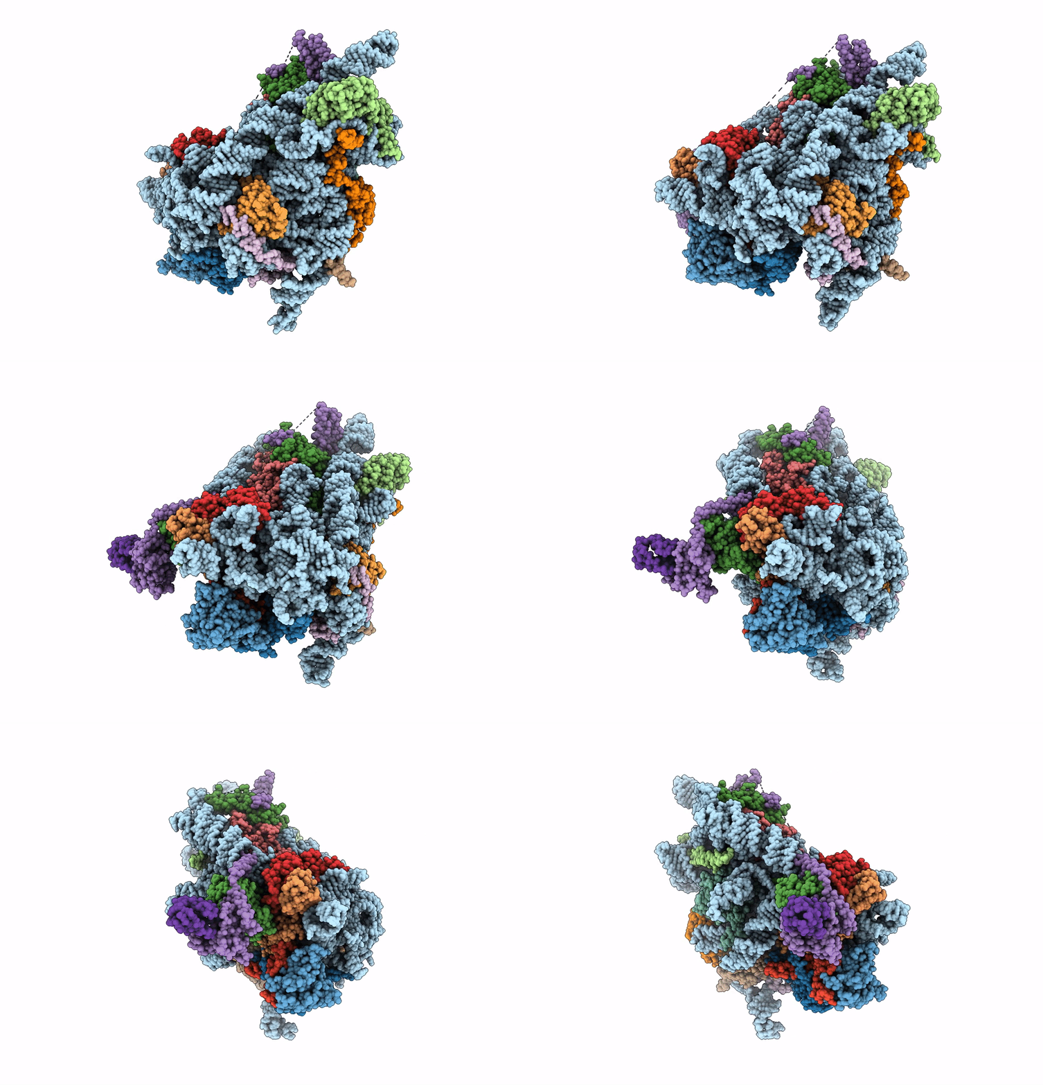
8C83 image
Entry Detail
PDB ID:
8C83
EMDB ID:
Keywords:
Title:
Cryo-EM structure of in vitro reconstituted Otu2-bound Ub-40S complex
Biological Source:
Source Organism:
Host Organism:
PDB Version:
Deposition Date:
2023-01-18
Release Date:
2023-05-24
Method Details:
Experimental Method:
Resolution:
3.00 Å
Aggregation State:
PARTICLE
Reconstruction Method:
SINGLE PARTICLE
Macromolecular Entities
Polymer Type:polyribonucleotide
Description:18S ribosomal RNA
Chain IDs:C (auth: 2)
Chain Length:1800
Number of Molecules:1
Biological Source:Saccharomyces cerevisiae
Polymer Type:polypeptide(L)
Description:40S ribosomal protein S0-A
Chain IDs:D (auth: P)
Chain Length:252
Number of Molecules:1
Biological Source:Saccharomyces cerevisiae
Polymer Type:polypeptide(L)
Description:40S ribosomal protein S2
Chain IDs:E (auth: R)
Chain Length:254
Number of Molecules:1
Biological Source:Saccharomyces cerevisiae
Polymer Type:polypeptide(L)
Description:40S ribosomal protein S4-A
Chain IDs:F (auth: S)
Chain Length:261
Number of Molecules:1
Biological Source:Saccharomyces cerevisiae
Polymer Type:polypeptide(L)
Description:40S ribosomal protein S6-A
Chain IDs:G (auth: T)
Chain Length:236
Number of Molecules:1
Biological Source:Saccharomyces cerevisiae
Polymer Type:polypeptide(L)
Description:40S ribosomal protein S7-A
Chain IDs:H (auth: U)
Chain Length:190
Number of Molecules:1
Biological Source:Saccharomyces cerevisiae
Polymer Type:polypeptide(L)
Description:40S ribosomal protein S8-B
Chain IDs:I (auth: V)
Chain Length:200
Number of Molecules:1
Biological Source:Saccharomyces cerevisiae
Polymer Type:polypeptide(L)
Description:40S ribosomal protein S9-A
Chain IDs:J (auth: W)
Chain Length:197
Number of Molecules:1
Biological Source:Saccharomyces cerevisiae
Polymer Type:polypeptide(L)
Description:40S ribosomal protein S11-A
Chain IDs:K (auth: X)
Chain Length:307
Number of Molecules:1
Biological Source:Saccharomyces cerevisiae
Polymer Type:polypeptide(L)
Description:40S ribosomal protein S13
Chain IDs:L (auth: Y)
Chain Length:151
Number of Molecules:1
Biological Source:Saccharomyces cerevisiae
Polymer Type:polypeptide(L)
Description:40S ribosomal protein S21-A
Chain IDs:M (auth: a)
Chain Length:87
Number of Molecules:1
Biological Source:Saccharomyces cerevisiae
Polymer Type:polypeptide(L)
Description:40S ribosomal protein S22-A
Chain IDs:N (auth: b)
Chain Length:130
Number of Molecules:1
Biological Source:Saccharomyces cerevisiae
Polymer Type:polypeptide(L)
Description:40S ribosomal protein S23-A
Chain IDs:O (auth: c)
Chain Length:145
Number of Molecules:1
Biological Source:Saccharomyces cerevisiae
Polymer Type:polypeptide(L)
Description:40S ribosomal protein S24-A
Chain IDs:P (auth: d)
Chain Length:135
Number of Molecules:1
Biological Source:Saccharomyces cerevisiae
Polymer Type:polypeptide(L)
Description:40S ribosomal protein S27-A
Chain IDs:Q (auth: f)
Chain Length:82
Number of Molecules:1
Biological Source:Saccharomyces cerevisiae
Polymer Type:polypeptide(L)
Description:40S ribosomal protein S30-A
Chain IDs:R (auth: g)
Chain Length:63
Number of Molecules:1
Biological Source:Saccharomyces cerevisiae
Polymer Type:polypeptide(L)
Description:OTU domain-containing protein 2
Mutations:C178S
Chain IDs:A (auth: x)
Chain Length:307
Number of Molecules:1
Biological Source:Saccharomyces cerevisiae
Polymer Type:polypeptide(L)
Description:Ubiquitin
Chain IDs:B (auth: y)
Chain Length:151
Number of Molecules:1
Biological Source:Saccharomyces cerevisiae
Ligand Molecules
Primary Citation
Molecular basis for recognition and deubiquitination of 40S ribosomes by Otu2.
Nat Commun 14 2730 2730 (2023)
PMID: 37169754 DOI: 10.1038/s41467-023-38161-w

Abstact

In actively translating 80S ribosomes the ribosomal protein eS7 of the 40S subunit is monoubiquitinated by the E3 ligase Not4 and deubiquitinated by Otu2 upon ribosomal subunit recycling. Despite its importance for translation efficiency the exact role and structural basis for this translational reset is poorly understood. Here, structural analysis by cryo-electron microscopy of native and reconstituted Otu2-bound ribosomal complexes reveals that Otu2 engages 40S subunits mainly between ribosome recycling and initiation stages. Otu2 binds to several sites on the intersubunit surface of the 40S that are not occupied by any other 40S-binding factors. This binding mode explains the discrimination against 80S ribosomes via the largely helical N-terminal domain of Otu2 as well as the specificity for mono-ubiquitinated eS7 on 40S. Collectively, this study reveals mechanistic insights into the Otu2-driven deubiquitination steps for translational reset during ribosome recycling/(re)initiation.

Legend

Protein

Chemical

Disease

Primary Citation of related structures