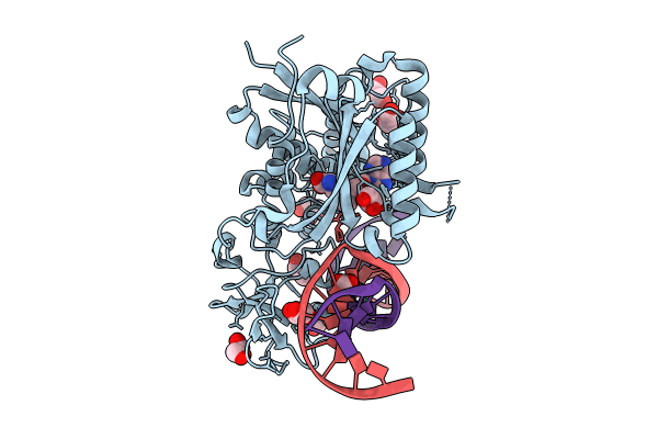
8C58 image
Entry Detail
PDB ID:
8C58
Keywords:
Title:
CpG specific M.MpeI methyltransferase crystallized in the presence of 5-hydroxycytosine and 5-methylcytosine containing dsDNA
Biological Source:
PDB Version:
Deposition Date:
2023-01-06
Release Date:
2024-01-17
Method Details:
Experimental Method:
Resolution:
1.85 Å
R-Value Free:
0.19
R-Value Work:
0.16
R-Value Observed:
0.16
Space Group:
P 41 21 2
Macromolecular Entities
Polymer Type:polypeptide(L)
Description:Cytosine-specific methyltransferase
Chain IDs:A
Chain Length:395
Number of Molecules:1
Biological Source:Malacoplasma penetrans HF-2
Polymer Type:polydeoxyribonucleotide
Description:DNA (5'-D(*CP*CP*AP*CP*AP*TP*GP*(5OC)P*GP*CP*TP*GP*AP*A)-3')
Chain IDs:B
Chain Length:14
Number of Molecules:1
Biological Source:synthetic construct
Polymer Type:polydeoxyribonucleotide
Description:DNA (5'-D(*GP*TP*TP*CP*AP*GP*(5CM)P*GP*CP*AP*TP*GP*TP*G)-3')
Chain IDs:C
Chain Length:14
Number of Molecules:1
Biological Source:synthetic construct
Primary Citation
Cytosine analogues as DNA methyltransferase substrates.
Nucleic Acids Res. 52 9267 9281 (2024)
PMID: 38966999 DOI: 10.1093/nar/gkae568

Abstact

DNA methyltransferases are drug targets for myelodysplastic syndrome (MDS), chronic myelomonocytic leukemia (CMML), acute myelogenous leukemia (AML) and possibly β-hemoglobinopathies. We characterize the interaction of nucleoside analogues in DNA with a prokaryotic CpG-specific DNA methyltransferase (M.MpeI) as a model for mammalian DNMT1 methyltransferases. We tested DNA containing 5-hydroxymethylcytosine (5hmC), 5-hydroxycytosine (5OHC), 5-methyl-2-pyrimidinone (in the ribosylated form known as 5-methylzebularine, 5mZ), 5,6-dihydro-5-azacytosine (dhaC), 5-fluorocytosine (5FC), 5-chlorocytosine (5ClC), 5-bromocytosine (5BrC) and 5-iodocytosine (5IC). Covalent complex formation was by far most efficient for 5FC. Non-covalent complexes were most abundant for dhaC and 5mZ. Surprisingly, we observed methylation of 5IC and 5BrC, and to a lesser extent 5ClC and 5FC, in the presence, but not the absence of small molecule thiol nucleophiles. For 5IC and 5BrC, we demonstrated by mass spectrometry that the reactions were due to methyltransferase driven dehalogenation, followed by methylation. Crystal structures of M.MpeI-DNA complexes capture the 'in' conformation of the active site loop for analogues with small or rotatable (5mZ) 5-substituents and its 'out' form for bulky 5-substituents. Since very similar 'in' and 'out' loop conformations were also observed for DNMT1, it is likely that our conclusions generalize to other DNA methyltransferases.

Legend

Protein

Chemical

Disease

Primary Citation of related structures