8C1N image
Entry Detail
PDB ID:
8C1N
Keywords:
Title:
FMDV 3D polymerase in complex with 3B1 protein solved in P212121 space group
Biological Source:
PDB Version:
Deposition Date:
2022-12-20
Release Date:
2023-04-26
Method Details:
Experimental Method:
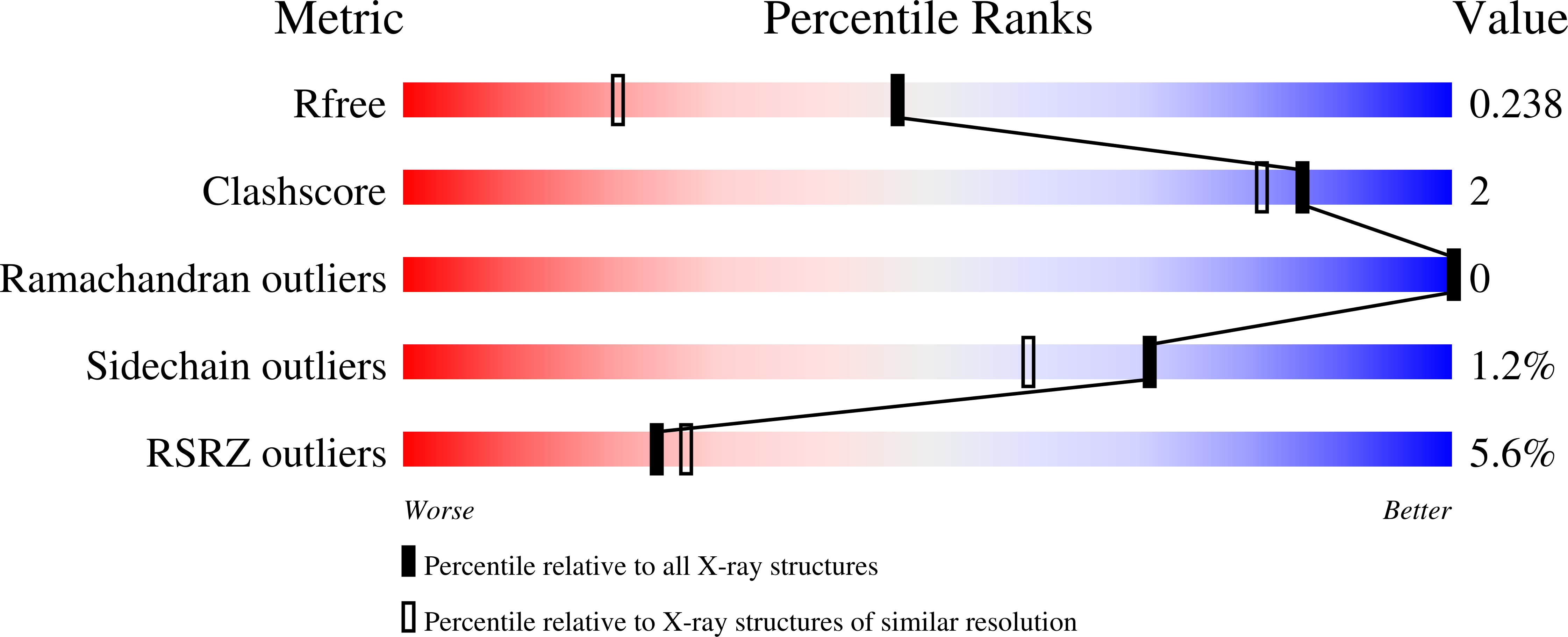
Resolution:
1.70 Å
R-Value Free:
0.23
R-Value Work:
0.21
R-Value Observed:
0.22
Space Group:
P 21 21 21
Macromolecular Entities
Polymer Type:polypeptide(L)
Description:RNA-directed RNA polymerase 3D-POL
Chain IDs:A, B
Chain Length:481
Number of Molecules:2
Biological Source:Foot-and-mouth disease virus
Polymer Type:polypeptide(L)
Description:Protein 3B-1
Chain IDs:C
Chain Length:23
Number of Molecules:1
Biological Source:Foot-and-mouth disease virus
Primary Citation
Dual role of the foot-and-mouth disease virus 3B1 protein in the replication complex: As protein primer and as an essential component to recruit 3Dpol to membranes.
Plos Pathog. 19 e1011373 e1011373 (2023)
PMID: 37126532 DOI: 10.1371/journal.ppat.1011373

Abstact

Picornavirus genome replication takes place in specialized intracellular membrane compartments that concentrate viral RNA and proteins as well as a number of host factors that also participate in the process. The core enzyme in the replication machinery is the viral RNA-dependent RNA polymerase (RdRP) 3Dpol. Replication requires the primer protein 3B (or VPg) attached to two uridine molecules. 3B uridylylation is also catalysed by 3Dpol. Another critical interaction in picornavirus replication is that between 3Dpol and the precursor 3AB, a membrane-binding protein responsible for the localization of 3Dpol to the membranous compartments at which replication occurs. Unlike other picornaviruses, the animal pathogen foot-and-mouth disease virus (FMDV), encodes three non-identical copies of the 3B (3B1, 3B2, and 3B3) that could be specialized in different functions within the replication complex. Here, we have used a combination of biophysics, molecular and structural biology approaches to characterize the functional binding of FMDV 3B1 to the base of the palm of 3Dpol. The 1.7 Å resolution crystal structure of the FMDV 3Dpol -3B1 complex shows that 3B1 simultaneously links two 3Dpol molecules by binding at the bottom of their palm subdomains in an almost symmetric way. The two 3B1 contact surfaces involve a combination of hydrophobic and basic residues at the N- (G5-P6, R9; Region I) and C-terminus (R16, L19-P20; Region II) of this small protein. Enzyme-Linked Immunosorbent Assays (ELISA) show that the two 3B1 binding sites play a role in 3Dpol binding, with region II presenting the highest affinity. ELISA assays show that 3Dpol has higher binding affinity for 3B1 than for 3B2 or 3B3. Membrane-based pull-down assays show that 3B1 region II, and to a lesser extent also region I play essential roles in mediating the interaction of 3AB with the polymerase and its recruitment to intracellular membranes.

Legend

Protein

Chemical

Disease

Primary Citation of related structures