8C0N image
Deposition Date 2022-12-19
Release Date 2023-03-08
Last Version Date 2023-11-15
Entry Detail
PDB ID:
8C0N
Title:
Crystal structure of the red form of the mTagFT fluorescent timer
Biological Source:
Source Organism:
Host Organism:
Method Details:
Experimental Method:
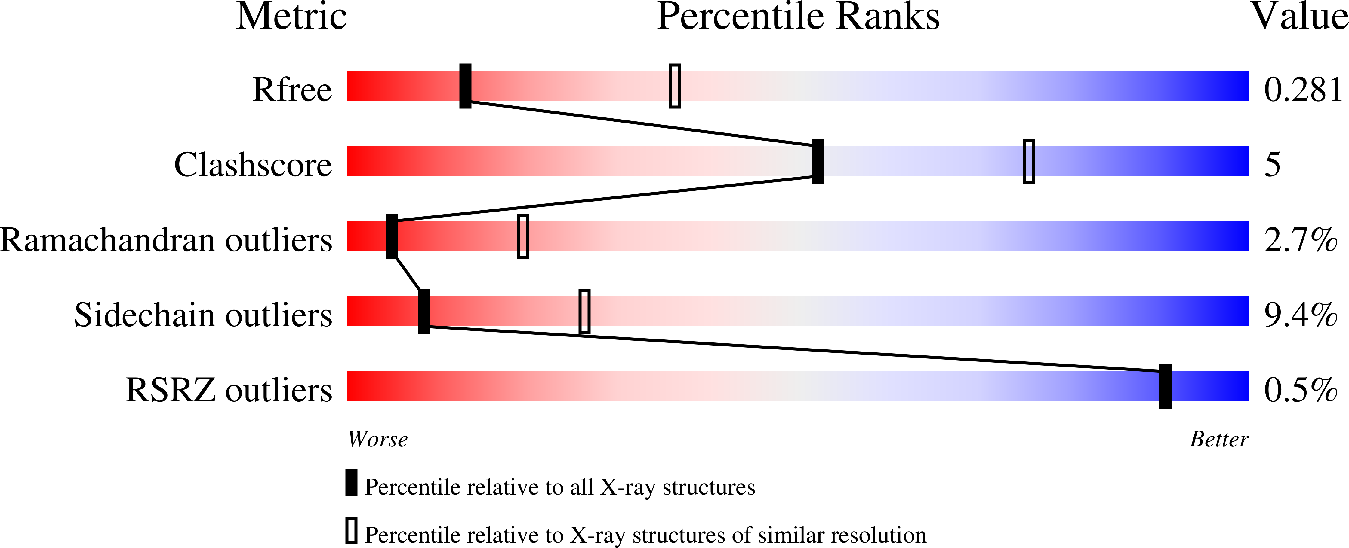
Resolution:
2.90 Å
R-Value Free:
0.27
R-Value Work:
0.24
R-Value Observed:
0.24
Space Group:
P 1 2 1
Macromolecular Entities
Polymer Type:polypeptide(L)
Molecule:Blue-to-red TagFT fluorescent timer
Chain IDs:A, B, C, D
Chain Length:277
Number of Molecules:4
Biological Source:Entacmaea quadricolor
Primary Citation
Blue-to-Red TagFT, mTagFT, mTsFT, and Green-to-FarRed mNeptusFT2 Proteins, Genetically Encoded True and Tandem Fluorescent Timers.
Int J Mol Sci 24 ? ? (2023)
PMID: 36834686 DOI: 10.3390/ijms24043279

Abstact

True genetically encoded monomeric fluorescent timers (tFTs) change their fluorescent color as a result of the complete transition of the blue form into the red form over time. Tandem FTs (tdFTs) change their color as a consequence of the fast and slow independent maturation of two forms with different colors. However, tFTs are limited to derivatives of the mCherry and mRuby red fluorescent proteins and have low brightness and photostability. The number of tdFTs is also limited, and there are no blue-to-red or green-to-far-red tdFTs. tFTs and tdFTs have not previously been directly compared. Here, we engineered novel blue-to-red tFTs, called TagFT and mTagFT, which were derived from the TagRFP protein. The main spectral and timing characteristics of the TagFT and mTagFT timers were determined in vitro. The brightnesses and photoconversions of the TagFT and mTagFT tFTs were characterized in live mammalian cells. The engineered split version of the TagFT timer matured in mammalian cells at 37 °C and allowed the detection of interactions between two proteins. The TagFT timer under the control of the minimal arc promoter, successfully visualized immediate-early gene induction in neuronal cultures. We also developed and optimized green-to-far-red and blue-to-red tdFTs, named mNeptusFT and mTsFT, which were based on mNeptune-sfGFP and mTagBFP2-mScarlet fusion proteins, respectively. We developed the FucciFT2 system based on the TagFT-hCdt1-100/mNeptusFT2-hGeminin combination, which could visualize the transitions between the G1 and S/G2/M phases of the cell cycle with better resolution than the conventional Fucci system because of the fluorescent color changes of the timers over time in different phases of the cell cycle. Finally, we determined the X-ray crystal structure of the mTagFT timer and analyzed it using directed mutagenesis.

Legend

Protein

Chemical

Disease

Primary Citation of related structures