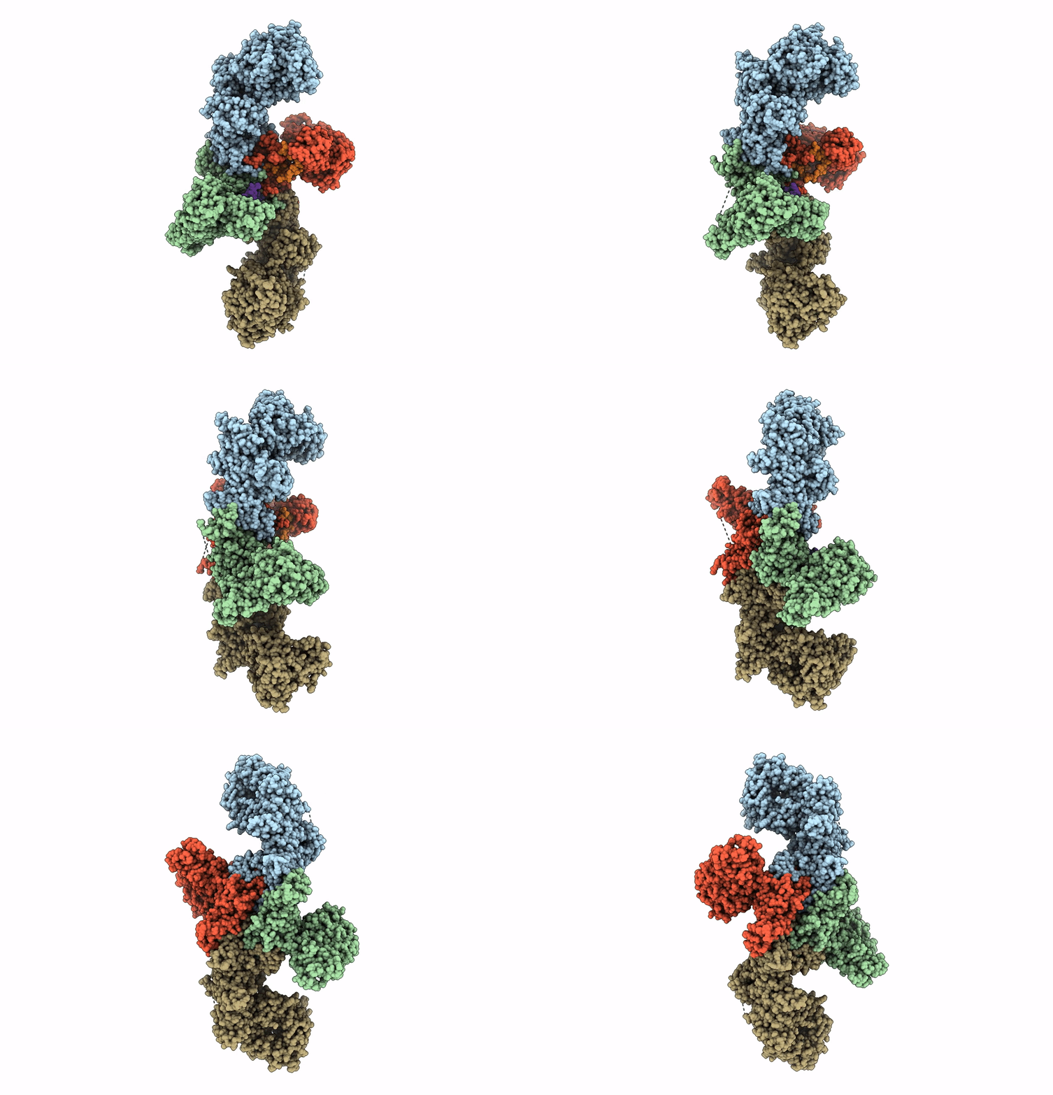
8C06 image
Deposition Date 2022-12-16
Release Date 2023-08-23
Last Version Date 2025-07-09
Entry Detail
PDB ID:
8C06
Keywords:
Title:
Structure of Dimeric HECT E3 Ubiquitin Ligase UBR5
Biological Source:
Source Organism:
Homo sapiens (Taxon ID: 9606)
Host Organism:
Method Details:
Experimental Method:
Resolution:
2.70 Å
Aggregation State:
PARTICLE
Reconstruction Method:
SINGLE PARTICLE
Macromolecular Entities
Polymer Type:polypeptide(L)
Molecule:E3 ubiquitin-protein ligase UBR5
Gene (Uniprot):UBR5
Mutations:K503R, L710D
Chain IDs:A, B, C (auth: D), D (auth: E), E (auth: F), F (auth: G)
Chain Length:2806
Number of Molecules:6
Biological Source:Homo sapiens
Ligand Molecules
Primary Citation
Structural snapshots along K48-linked ubiquitin chain formation by the HECT E3 UBR5.
Nat.Chem.Biol. 20 190 200 (2024)
PMID: 37620400 DOI: 10.1038/s41589-023-01414-2

Abstact

Ubiquitin (Ub) chain formation by homologous to E6AP C-terminus (HECT)-family E3 ligases regulates vast biology, yet the structural mechanisms remain unknown. We used chemistry and cryo-electron microscopy (cryo-EM) to visualize stable mimics of the intermediates along K48-linked Ub chain formation by the human E3, UBR5. The structural data reveal a ≈ 620 kDa UBR5 dimer as the functional unit, comprising a scaffold with flexibly tethered Ub-associated (UBA) domains, and elaborately arranged HECT domains. Chains are forged by a UBA domain capturing an acceptor Ub, with its K48 lured into the active site by numerous interactions between the acceptor Ub, manifold UBR5 elements and the donor Ub. The cryo-EM reconstructions allow defining conserved HECT domain conformations catalyzing Ub transfer from E2 to E3 and from E3. Our data show how a full-length E3, ubiquitins to be adjoined, E2 and intermediary products guide a feed-forward HECT domain conformational cycle establishing a highly efficient, broadly targeting, K48-linked Ub chain forging machine.

Legend

Protein

Chemical

Disease

Primary Citation of related structures