8BWT image
Deposition Date 2022-12-07
Release Date 2023-08-30
Last Version Date 2024-07-03
Entry Detail
PDB ID:
8BWT
Keywords:
Title:
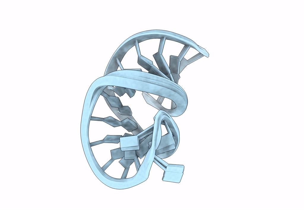
Structure of a symmetrical internal loop motif with three consecutive U:U mismatches from stem-loop 1 in the 3'-UTR of the SARS-CoV2 genomic RNA
Biological Source:
Method Details:
Experimental Method:
Conformers Calculated:
100
Conformers Submitted:
10
Selection Criteria:
structures with the lowest energy
Macromolecular Entities
Polymer Type:polyribonucleotide
Molecule:RNA (26-MER)
Chain IDs:A
Chain Length:26
Number of Molecules:1
Biological Source:Severe acute respiratory syndrome coronavirus 2
Ligand Molecules
Primary Citation
Structure of an internal loop motif with three consecutive U•U mismatches from stem-loop 1 in the 3'-UTR of the SARS-CoV-2 genomic RNA.
Nucleic Acids Res. 52 6687 6706 (2024)
PMID: 38783391 DOI: 10.1093/nar/gkae349

Abstact

The single-stranded RNA genome of SARS-CoV-2 is highly structured. Numerous helical stem-loop structures interrupted by mismatch motifs are present in the functionally important 5'- and 3'-UTRs. These mismatches modulate local helical geometries and feature unusual arrays of hydrogen bonding donor and acceptor groups. However, their conformational and dynamical properties cannot be directly inferred from chemical probing and are difficult to predict theoretically. A mismatch motif (SL1-motif) consisting of three consecutive U•U base pairs is located in stem-loop 1 of the 3'-UTR. We combined NMR-spectroscopy and MD-simulations to investigate its structure and dynamics. All three U•U base pairs feature two direct hydrogen bonds and are as stable as Watson-Crick A:U base pairs. Plasmodium falciparum 25S rRNA contains a triple U•U mismatch motif (Pf-motif) differing from SL1-motif only with respect to the orientation of the two closing base pairs. Interestingly, while the geometry of the outer two U•U mismatches was identical in both motifs the preferred orientation of the central U•U mismatch was different. MD simulations and potassium ion titrations revealed that the potassium ion-binding mode to the major groove is connected to the different preferred geometries of the central base pair in the two motifs.

Legend

Protein

Chemical

Disease

Primary Citation of related structures