8BW0 image
Deposition Date 2022-12-06
Release Date 2024-01-24
Last Version Date 2024-11-13
Entry Detail
PDB ID:
8BW0
Keywords:
Title:
Structure of CEACAM5 A3-B3 domain in Complex with Tusamitamab Fab
Biological Source:
Source Organism:
Mus sp. (Taxon ID: 10095)
Homo sapiens (Taxon ID: 9606)
Host Organism:
Method Details:
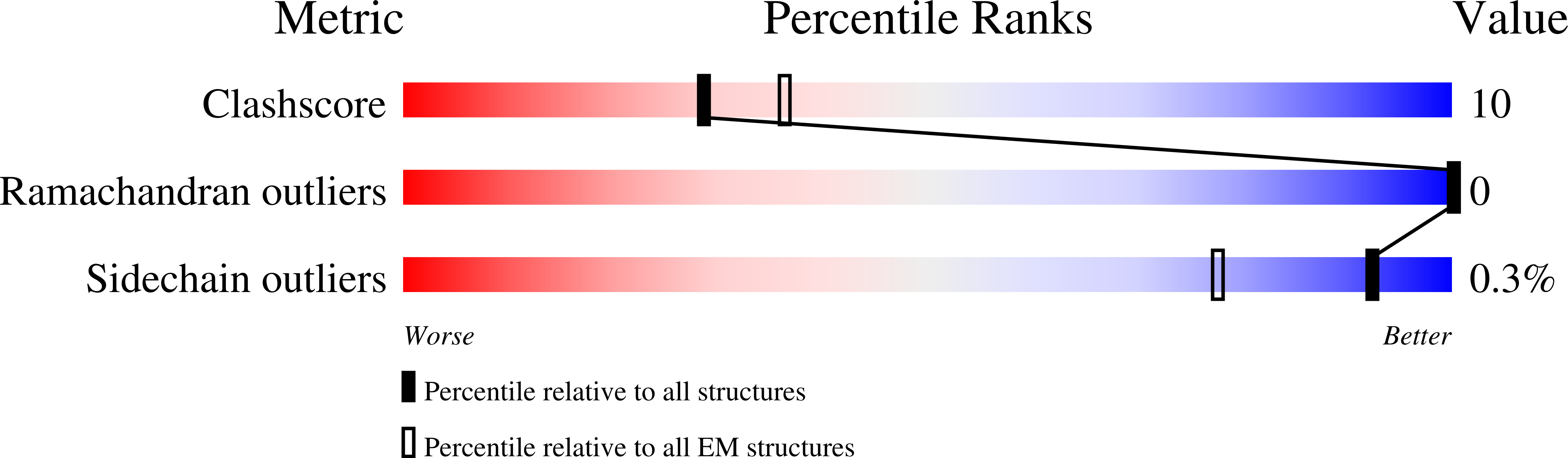
Experimental Method:
Resolution:
3.11 Å
Aggregation State:
PARTICLE
Reconstruction Method:
SINGLE PARTICLE
Macromolecular Entities
Structures with similar UniProt ID
Protein Blast
Polymer Type:polypeptide(L)
Molecule:Carcinoembryonic antigen-related cell adhesion molecule 5
Gene (Uniprot):CEACAM5
Chain IDs:C
Chain Length:193
Number of Molecules:1
Biological Source:Homo sapiens
Polymer Type:polypeptide(L)
Molecule:Tusamitamab Fab heavy Chain
Chain IDs:A (auth: H)
Chain Length:234
Number of Molecules:1
Biological Source:Mus sp.
Polymer Type:polypeptide(L)
Molecule:Tusamitamab Light Chain
Chain IDs:B (auth: L)
Chain Length:214
Number of Molecules:1
Biological Source:Mus sp.
Ligand Molecules
Primary Citation
Structural insights into epitope-paratope interactions of a monoclonal antibody targeting CEACAM5-expressing tumors.
Nat Commun 15 9377 9377 (2024)
PMID: 39477960 DOI: 10.1038/s41467-024-53746-9

Abstact

Carcinoembryonic antigen-related cell adhesion molecules (CEACAMs) are overexpressed in some tumor types. The antibody-drug conjugate tusamitamab ravtansine specifically recognizes the A3-B3 domains of human CEACAM5 (hCEACAM5). To understand this specificity, here we map the epitope-paratope interface between the A3-B3 domains of hCEACAM5 (hCEACAM5A3-B3) and the antigen-binding fragment of tusamitamab (tusa Fab). We use hydrogen/deuterium exchange mass spectrometry to identify the tusa Fab paratope, which involves heavy chain (HC) residues 101-109 and light chain residues 48-54 and 88-104. Using surface plasmon resonance, we demonstrate that alanine variants of HC residues 96-108 abolish binding to hCEACAM5, suggesting that these residues are critical for tusa-Fab-antigen complex formation. The cryogenic electron microscopy structure of the hCEACAM5A3-B3- tusa Fab complex (3.11 Å overall resolution) reveals a discontinuous epitope involving residues in the A3-B3 domains and an N-linked mannose at residue Asn612. Conformational constraints on the epitope-paratope interface enable tusamitamab to target hCEACAM5A3-B3 and distinguish CEACAM5 from other CEACAMs.

Legend

Protein

Chemical

Disease

Primary Citation of related structures