8BVI image
Deposition Date 2022-12-04
Release Date 2023-12-13
Last Version Date 2025-07-09
Entry Detail
PDB ID:
8BVI
Keywords:
Title:
Crystal structure of the METTL9-like histidine methyltransferase from Ostreococcus tauri
Biological Source:
Source Organism:
Method Details:
Experimental Method:
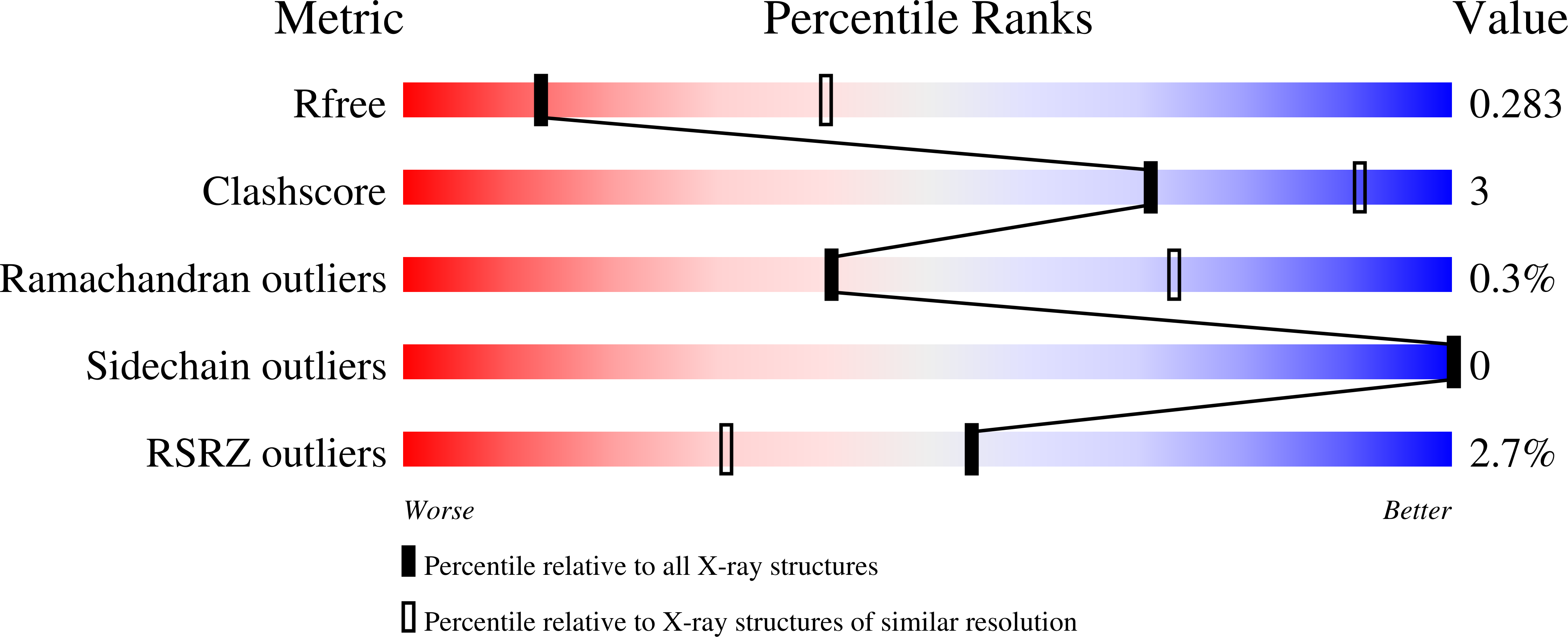
Resolution:
3.10 Å
R-Value Free:
0.28
R-Value Work:
0.22
R-Value Observed:
0.22
Space Group:
P 21 3
Macromolecular Entities
Polymer Type:polypeptide(L)
Molecule:DREV methyltransferase
Gene (Uniprot):OT_ostta03g04640
Chain IDs:A
Chain Length:310
Number of Molecules:1
Biological Source:Ostreococcus tauri
Primary Citation
Orthologues of the human protein histidine methyltransferase METTL9 display distinct substrate specificities.
J.Biol.Chem. ? 110318 110318 (2025)
PMID: 40451431 DOI: 10.1016/j.jbc.2025.110318

Abstact

The human (Homo sapiens; Hs) methyltransferase (MTase) METTL9 is the first enzyme shown to generate 1-methylhistidine (π-methylhistidine) in proteins. METTL9 preferentially methylates an alternating histidine (HxH) motif, where "x" is a small, uncharged amino acid, and multiple substrates have been identified. Putative METTL9 orthologues are found in most eukaryotes, and we have here investigated the activity of such enzymes from several species, representing all five eukaryotic supergroups. The majority of the tested enzymes demonstrated in vitro MTase activity on the prototype HsMETTL9 substrates ARMC6 and DNAJB12. We also detected protein methylation activity of the Caenorhabditis elegans METTL9 which had previously been suggested to be a DNA MTase. However, METTL9 from the fruit fly (Drosophila melanogaster; Dm) and the picoplankton Ostreococcus tauri (Ot) displayed distinct substrate specificities, differing from each other and from that of HsMETTL9. These differences were observed when recombinant proteins and short peptides were used as METTL9 substrates. To further analyze substrate specificity, we used peptide arrays to systematically replace the "x" residue and the residues flanking the HxH motif in a substrate peptide. This revealed varying degrees of tolerance among the METTL9 orthologues (Hs > Dm > Ot) for substitutions at these positions. Our results show that the METTL9 orthologues, although requiring an HxH target site, have evolved different substrate specificities, likely due to differing biologically relevant substrates in the respective organisms. Furthermore, we solved the X-ray structure of OtMETTL9, revealing several differences from the previously published HsMETTL9 structures that may explain its distinct substrate specificity.

Legend

Protein

Chemical

Disease

Primary Citation of related structures