8BT1 image
Deposition Date 2022-11-27
Release Date 2023-02-22
Last Version Date 2024-06-19
Entry Detail
PDB ID:
8BT1
Title:
YdaT transcription regulator (CII functional analog)
Biological Source:
Source Organism:
Method Details:
Experimental Method:
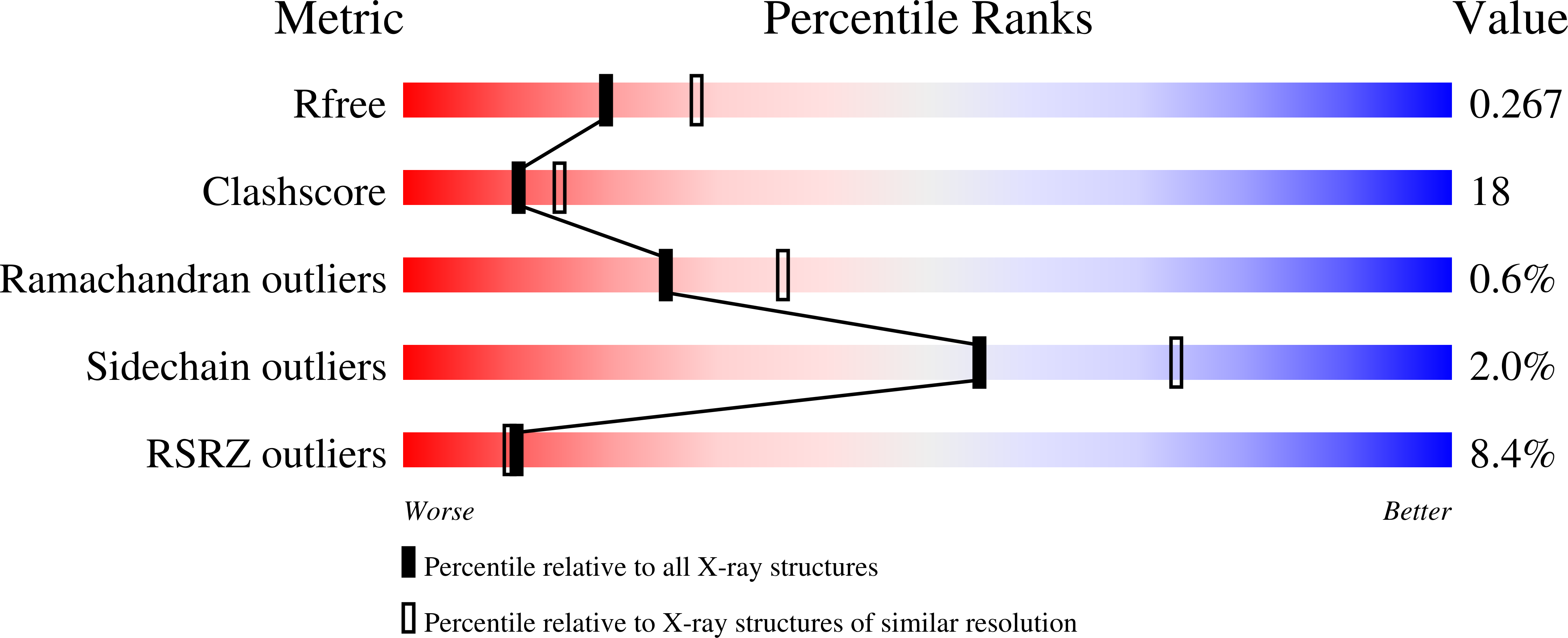
Resolution:
2.40 Å
R-Value Free:
0.26
R-Value Work:
0.21
R-Value Observed:
0.21
Space Group:
P 1
Macromolecular Entities
Polymer Type:polypeptide(L)
Molecule:YdaT_toxin domain-containing protein
Chain IDs:A, B, C, D
Chain Length:162
Number of Molecules:4
Biological Source:Escherichia coli O157:H7
Primary Citation
Structural basis of DNA binding by YdaT, a functional equivalent of the CII repressor in the cryptic prophage CP-933P from Escherichia coli O157:H7.
Acta Crystallogr D Struct Biol 79 245 258 (2023)
PMID: 36876434 DOI: 10.1107/S2059798323001249

Abstact

YdaT is a functional equivalent of the CII repressor in certain lambdoid phages and prophages. YdaT from the cryptic prophage CP-933P in the genome of Escherichia coli O157:H7 is functional as a DNA-binding protein and recognizes a 5'-TTGATTN6AATCAA-3' inverted repeat. The DNA-binding domain is a helix-turn-helix (HTH)-containing POU domain and is followed by a long α-helix (α6) that forms an antiparallel four-helix bundle, creating a tetramer. The loop between helix α2 and the recognition helix α3 in the HTH motif is unusually long compared with typical HTH motifs, and is highly variable in sequence and length within the YdaT family. The POU domains have a large degree of freedom to move relative to the helix bundle in the free structure, but their orientation becomes fixed upon DNA binding.

Legend

Protein

Chemical

Disease

Primary Citation of related structures