
Deposition Date
2022-10-28
Release Date
2023-05-10
Last Version Date
2024-05-01
Entry Detail
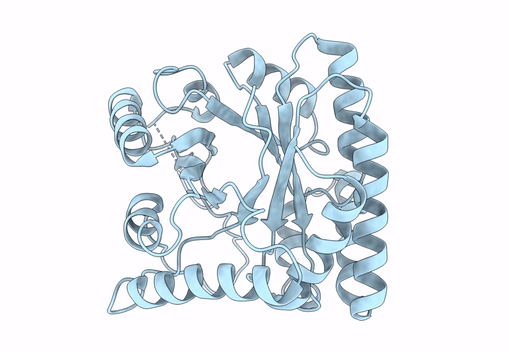
PDB ID:
8BGR
Keywords:
Title:
FAD-independent Methylene-Tetrahydrofolate Reductase from Mycobacterium hassiacum
Biological Source:
Source Organism(s):
Mycolicibacterium hassiacum (Taxon ID: 46351)
Expression System(s):
Method Details:
Experimental Method:
Resolution:
1.80 Å
R-Value Free:
0.24
R-Value Work:
0.23
R-Value Observed:
0.23
Space Group:
P 43 21 2
