Deposition Date
2022-10-28
Release Date
2023-02-15
Last Version Date
2024-10-16
Entry Detail
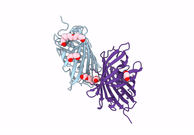
Biological Source:
Source Organism(s):
Discosoma sp. (Taxon ID: 86600)
Expression System(s):
Method Details:
Experimental Method:
Resolution:
2.00 Å
R-Value Free:
0.23
R-Value Work:
0.18
R-Value Observed:
0.19
Space Group:
I 1 2 1