Deposition Date
2022-10-27
Release Date
2023-11-08
Last Version Date
2025-05-21
Entry Detail
PDB ID:
8BGB
Keywords:
Title:
Structure of the citrate-bound extracytoplasmic PAS domain of histidine kinase CitA from Geobacillus thermodenitrificans
Biological Source:
Source Organism(s):
Geobacillus thermodenitrificans (Taxon ID: 33940)
Expression System(s):
Method Details:
Experimental Method:
Resolution:
1.70 Å
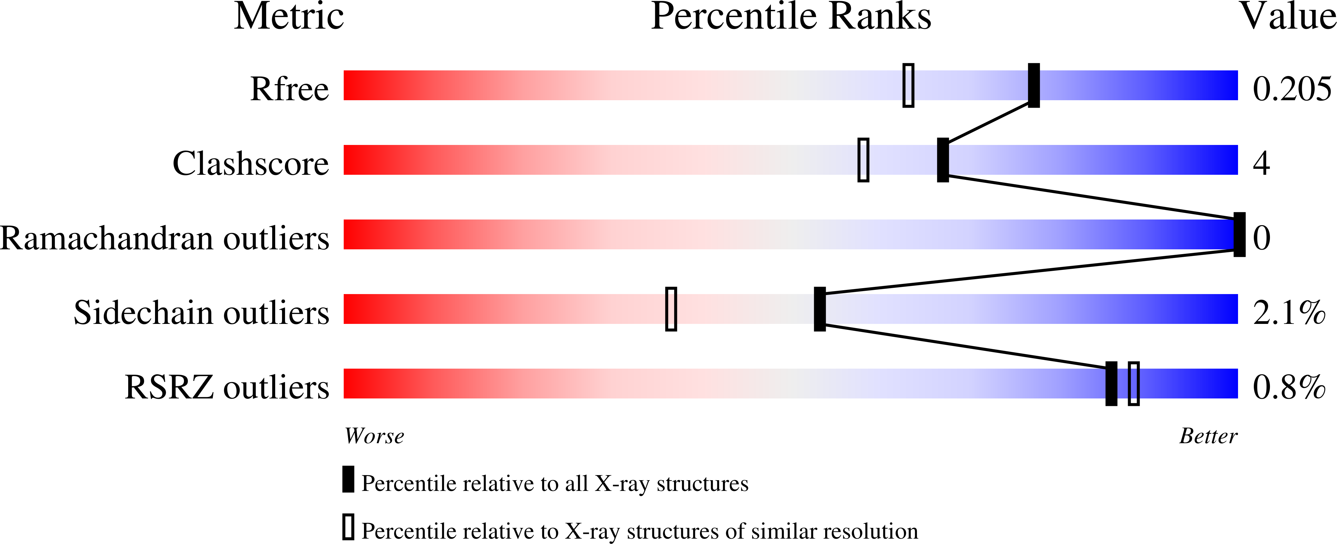
R-Value Free:
0.19
R-Value Work:
0.16
Space Group:
P 1 21 1