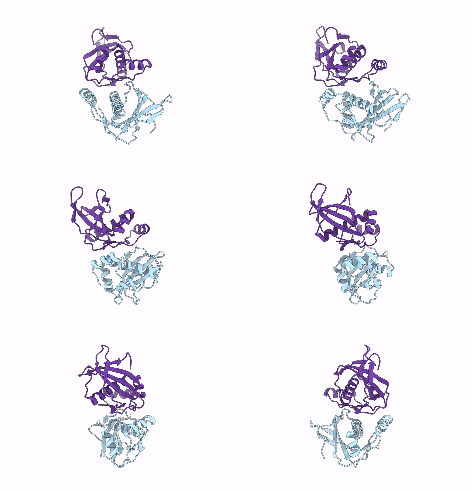
8BDU image
Deposition Date 2022-10-20
Release Date 2023-08-16
Last Version Date 2023-08-23
Entry Detail
PDB ID:
8BDU
Keywords:
Title:
H33 variant of DoBi scaffold based on PIH1D1 N-terminal domain
Biological Source:
Source Organism:
Homo sapiens (Taxon ID: 9606)
Method Details:
Experimental Method:
Resolution:
2.47 Å
R-Value Free:
0.25
R-Value Work:
0.21
R-Value Observed:
0.22
Space Group:
I 41 2 2
Macromolecular Entities
Structures with similar UniProt ID
Protein Blast
Polymer Type:polypeptide(L)
Molecule:PIH1 domain-containing protein 1
Gene (Uniprot):PIH1D1
Chain IDs:A, B
Chain Length:152
Number of Molecules:2
Biological Source:Homo sapiens
Ligand Molecules
Primary Citation
Diffraction anisotropy and paired refinement: crystal structure of H33, a protein binder to interleukin 10.
J.Appl.Crystallogr. 56 1261 1266 (2023)
PMID: 37555209 DOI: 10.1107/S160057672300479X

Abstact

Binder H33 is a small protein binder engineered by ribosome display to bind human interleukin 10. Crystals of binder H33 display severe diffraction anisotropy. A set of data files with correction for diffraction anisotropy based on different local signal-to-noise ratios was prepared. Paired refinement was used to find the optimal anisotropic high-resolution diffraction limit of the data: 3.13-2.47 Å. The structure of binder H33 belongs to the 2% of crystal structures with the highest solvent content in the Protein Data Bank.

Legend

Protein

Chemical

Disease

Primary Citation of related structures