8BCC image
Deposition Date 2022-10-15
Release Date 2023-03-08
Last Version Date 2024-02-07
Entry Detail
PDB ID:
8BCC
Keywords:
Title:
Human Brr2 Helicase Region in complex with C-tail deleted Jab1 and compound 39
Biological Source:
Source Organism:
Homo sapiens (Taxon ID: 9606)
Host Organism:
Method Details:
Experimental Method:
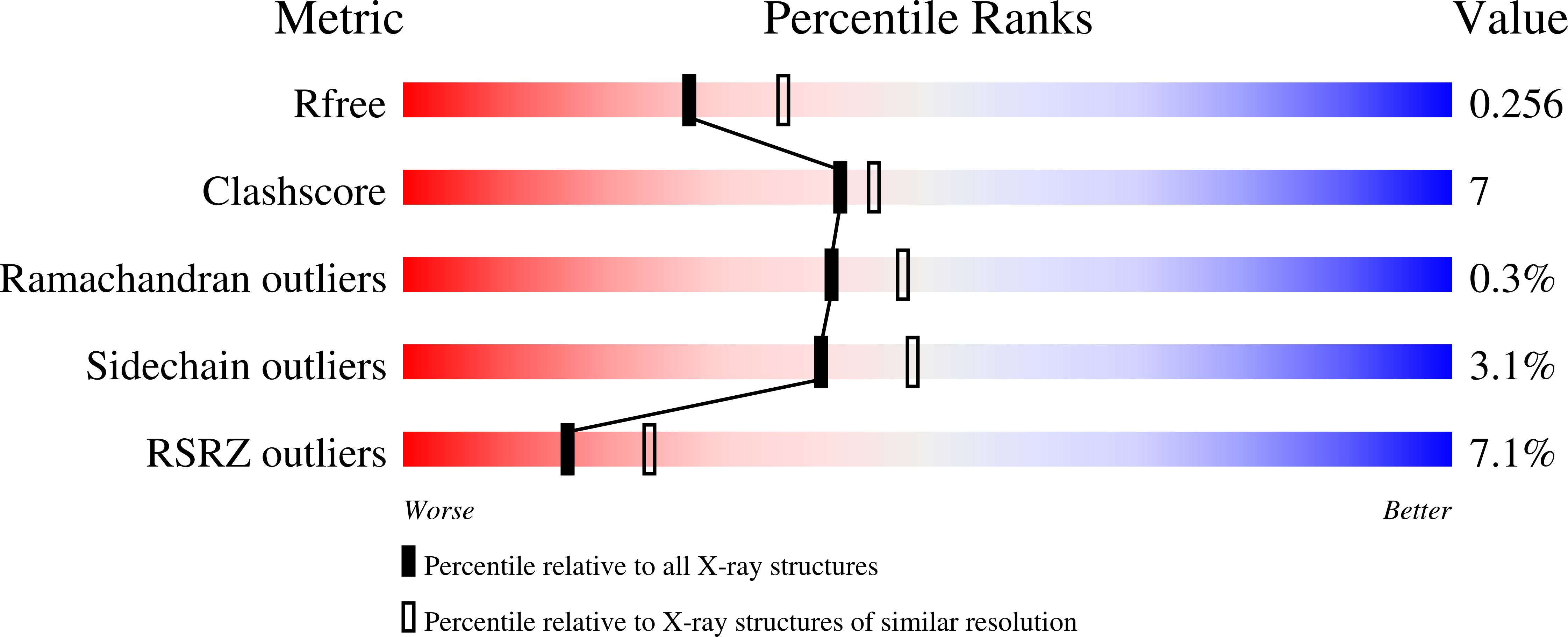
Resolution:
2.35 Å
R-Value Free:
0.25
R-Value Work:
0.20
R-Value Observed:
0.21
Space Group:
P 21 21 21
Macromolecular Entities
Polymer Type:polypeptide(L)
Molecule:U5 small nuclear ribonucleoprotein 200 kDa helicase
Gene (Uniprot):SNRNP200
Chain IDs:A (auth: B)
Chain Length:1747
Number of Molecules:1
Biological Source:Homo sapiens
Polymer Type:polypeptide(L)
Molecule:Pre-mRNA-processing-splicing factor 8
Gene (Uniprot):PRPF8
Chain IDs:B (auth: J)
Chain Length:263
Number of Molecules:1
Biological Source:Homo sapiens
Primary Citation
Conformation-dependent ligand hot spots in the spliceosomal RNA helicase BRR2.
Acta Crystallogr D Struct Biol 79 304 317 (2023)
PMID: 36974964 DOI: 10.1107/S2059798323001778

Abstact

The conversion of hits to leads in drug discovery involves the elaboration of chemical core structures to increase their potency. In fragment-based drug discovery, low-molecular-weight compounds are tested for protein binding and are subsequently modified, with the tacit assumption that the binding mode of the original hit will be conserved among the derivatives. However, deviations from binding mode conservation are rather frequently observed, but potential causes of these alterations remain incompletely understood. Here, two crystal forms of the spliceosomal RNA helicase BRR2 were employed as a test case to explore the consequences of conformational changes in the target protein on the binding behaviour of fragment derivatives. The initial fragment, sulfaguanidine, bound at the interface between the two helicase cassettes of BRR2 in one crystal form. Second-generation compounds devised by structure-guided docking were probed for their binding to BRR2 in a second crystal form, in which the original fragment-binding site was altered due to a conformational change. While some of the second-generation compounds retained binding to parts of the original site, others changed to different binding pockets of the protein. A structural bioinformatics analysis revealed that the fragment-binding sites correspond to predicted binding hot spots, which strongly depend on the protein conformation. This case study offers an example of extensive binding-mode changes during hit derivatization, which are likely to occur as a consequence of multiple binding hot spots, some of which are sensitive to the flexibility of the protein.

Legend

Protein

Chemical

Disease

Primary Citation of related structures