8BAQ image
Deposition Date 2022-10-11
Release Date 2023-07-12
Last Version Date 2024-02-07
Entry Detail
PDB ID:
8BAQ
Keywords:
Title:
E. coli C7 DarT1 in complex with NAD+
Biological Source:
Source Organism:
Escherichia coli (Taxon ID: 562)
Method Details:
Experimental Method:
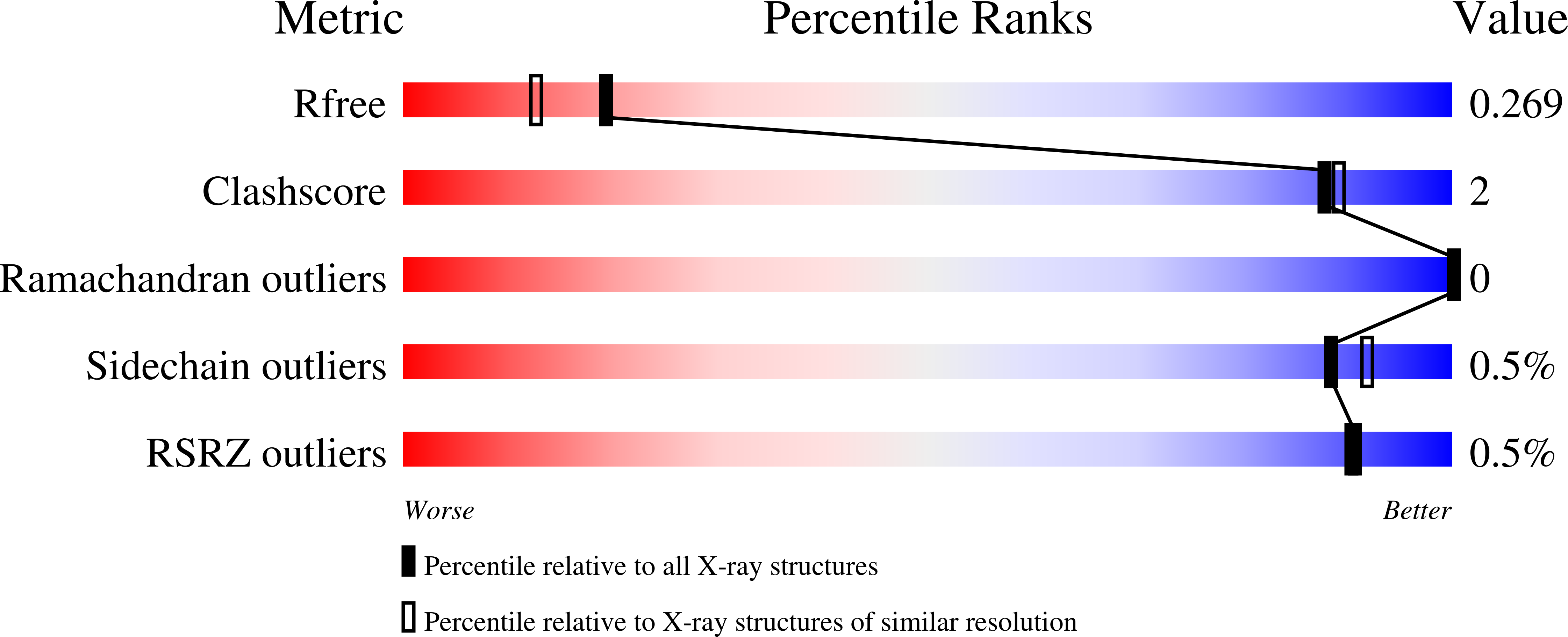
Resolution:
2.00 Å
R-Value Free:
0.24
R-Value Work:
0.19
Space Group:
P 31 2 1
Macromolecular Entities
Structures with similar UniProt ID
Protein Blast
Polymer Type:polypeptide(L)
Molecule:DarT ssDNA thymidine ADP-ribosyltransferase family protein
Mutagens:E152A
Chain IDs:A
Chain Length:208
Number of Molecules:1
Biological Source:Escherichia coli
Primary Citation
Molecular basis for the reversible ADP-ribosylation of guanosine bases.
Mol.Cell 83 2303 ? (2023)
PMID: 37390817 DOI: 10.1016/j.molcel.2023.06.013

Abstact

Modification of nucleic acids by ADP-ribosylation is catalyzed by various ADP-ribosyltransferases, including the DarT enzyme. The latter is part of the bacterial toxin-antitoxin (TA) system DarTG, which was shown to provide control of DNA replication and bacterial growth as well as protection against bacteriophages. Two subfamilies have been identified, DarTG1 and DarTG2, which are distinguished by their associated antitoxins. While DarTG2 catalyzes reversible ADP-ribosylation of thymidine bases employing a macrodomain as antitoxin, the DNA ADP-ribosylation activity of DarTG1 and the biochemical function of its antitoxin, a NADAR domain, are as yet unknown. Using structural and biochemical approaches, we show that DarT1-NADAR is a TA system for reversible ADP-ribosylation of guanosine bases. DarT1 evolved the ability to link ADP-ribose to the guanine amino group, which is specifically hydrolyzed by NADAR. We show that guanine de-ADP-ribosylation is also conserved among eukaryotic and non-DarT-associated NADAR members, indicating a wide distribution of reversible guanine modifications beyond DarTG systems.

Legend

Protein

Chemical

Disease

Primary Citation of related structures