8B6R image
Entry Detail
PDB ID:
8B6R
Keywords:
Title:
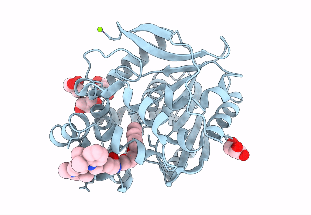
X-ray structure of the haloalkane dehalogenase HaloTag7 labeled with a chloroalkane Cyanine3 fluorophore substrate
Biological Source:
Source Organism:
Host Organism:
PDB Version:
Deposition Date:
2022-09-27
Release Date:
2023-07-26
Method Details:
Experimental Method:
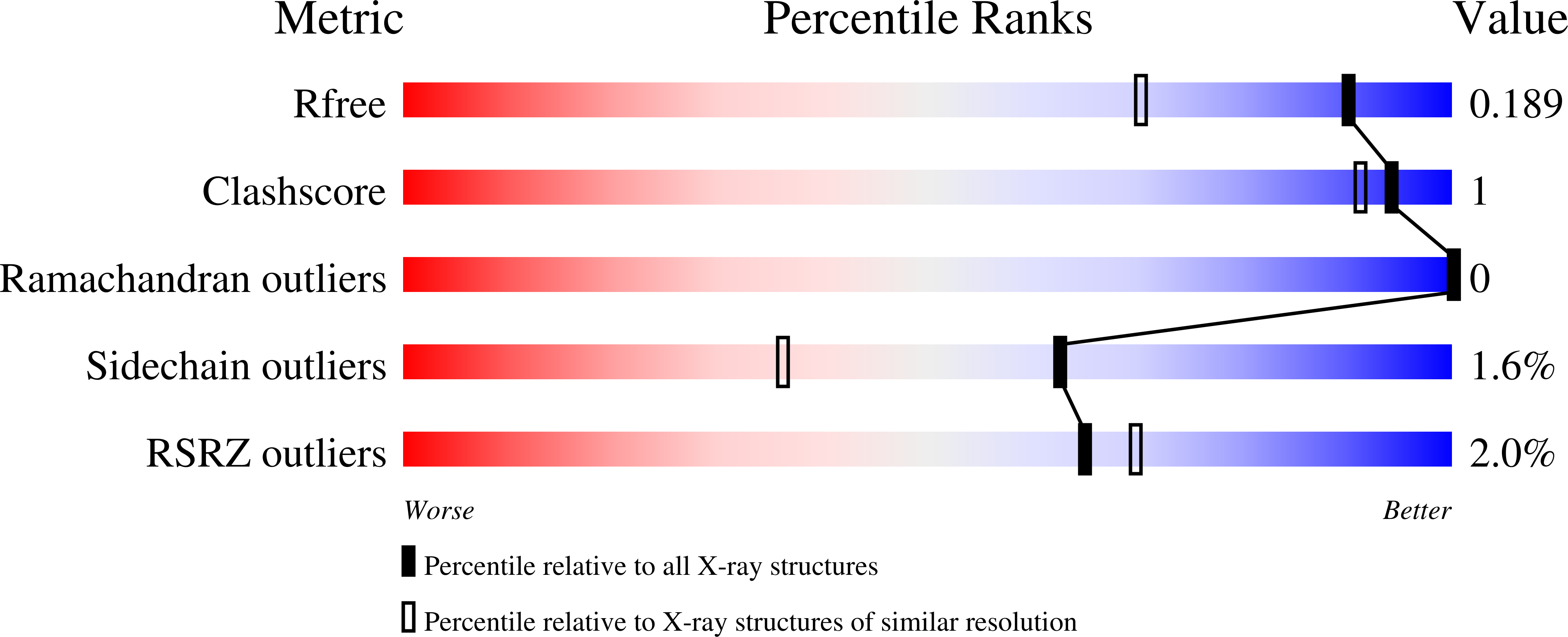
Resolution:
1.50 Å
R-Value Free:
0.19
R-Value Work:
0.16
R-Value Observed:
0.16
Space Group:
P 42 21 2
Macromolecular Entities
Polymer Type:polypeptide(L)
Description:Haloalkane dehalogenase
Chain IDs:A
Chain Length:293
Number of Molecules:1
Biological Source:Rhodococcus sp.
Primary Citation
A general method for the development of multicolor biosensors with large dynamic ranges.
Nat.Chem.Biol. 19 1147 1157 (2023)
PMID: 37291200 DOI: 10.1038/s41589-023-01350-1

Abstact

Fluorescent biosensors enable the study of cell physiology with spatiotemporal resolution; yet, most biosensors suffer from relatively low dynamic ranges. Here, we introduce a family of designed Förster resonance energy transfer (FRET) pairs with near-quantitative FRET efficiencies based on the reversible interaction of fluorescent proteins with a fluorescently labeled HaloTag. These FRET pairs enabled the straightforward design of biosensors for calcium, ATP and NAD+ with unprecedented dynamic ranges. The color of each of these biosensors can be readily tuned by changing either the fluorescent protein or the synthetic fluorophore, which enables simultaneous monitoring of free NAD+ in different subcellular compartments following genotoxic stress. Minimal modifications of these biosensors furthermore allow their readout to be switched to fluorescence intensity, fluorescence lifetime or bioluminescence. These FRET pairs thus establish a new concept for the development of highly sensitive and tunable biosensors.

Legend

Protein

Chemical

Disease

Primary Citation of related structures