8B02 image
Deposition Date 2022-09-07
Release Date 2022-11-30
Last Version Date 2024-01-31
Entry Detail
PDB ID:
8B02
Title:
Crystal structure of the dsRBD domain of tRNA-dihydrouridine(20) synthase from Amphimedon queenslandica
Biological Source:
Source Organism(s):
Expression System(s):
Method Details:
Experimental Method:
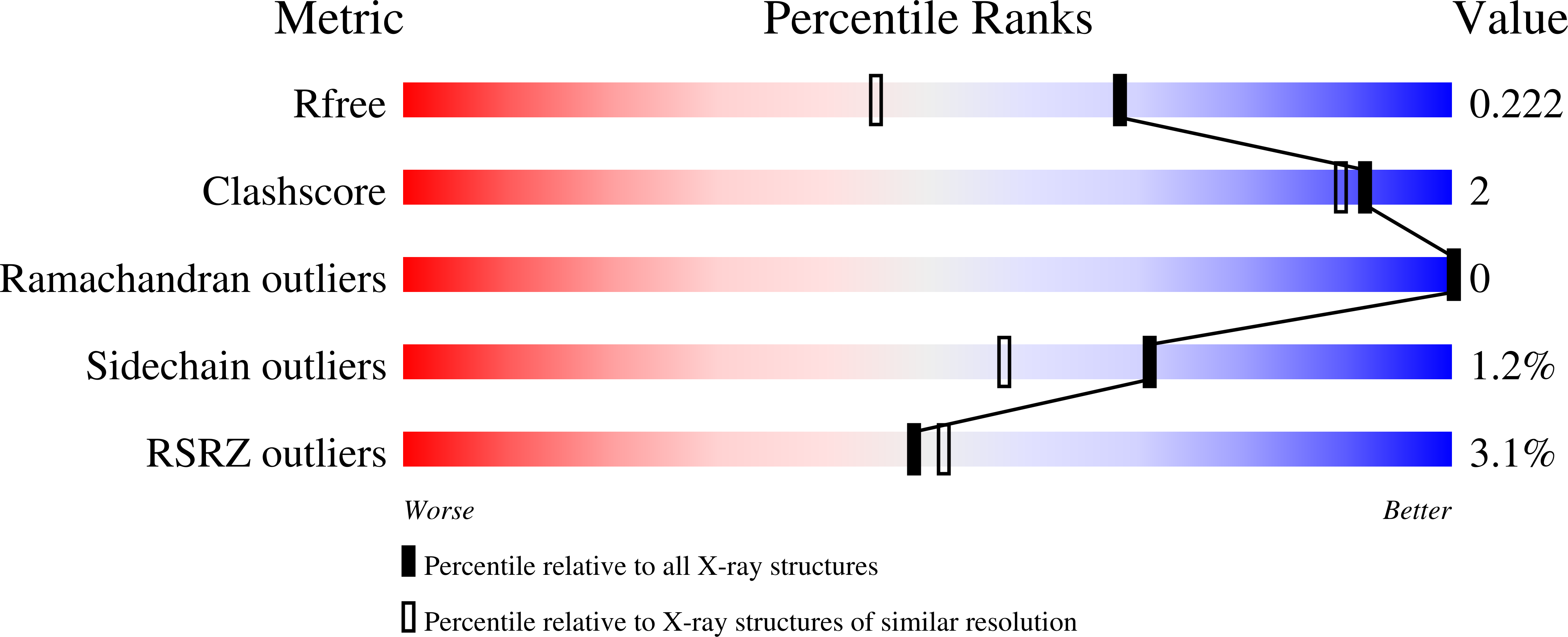
Resolution:
1.68 Å
R-Value Free:
0.22
R-Value Work:
0.20
R-Value Observed:
0.20
Space Group:
P 1 21 1
Macromolecular Entities
Structures with similar UniProt ID
Protein Blast
Polymer Type:polypeptide(L)
Molecule:DRBM domain-containing protein
Gene (Uniprot):100640958
Chain IDs:A, B
Chain Length:113
Number of Molecules:2
Biological Source:Amphimedon queenslandica
Primary Citation
Evolutionary Diversity of Dus2 Enzymes Reveals Novel Structural and Functional Features among Members of the RNA Dihydrouridine Synthases Family.
Biomolecules 12 ? ? (2022)
PMID: 36551188 DOI: 10.3390/biom12121760

Abstact

Dihydrouridine (D) is an abundant modified base found in the tRNAs of most living organisms and was recently detected in eukaryotic mRNAs. This base confers significant conformational plasticity to RNA molecules. The dihydrouridine biosynthetic reaction is catalyzed by a large family of flavoenzymes, the dihydrouridine synthases (Dus). So far, only bacterial Dus enzymes and their complexes with tRNAs have been structurally characterized. Understanding the structure-function relationships of eukaryotic Dus proteins has been hampered by the paucity of structural data. Here, we combined extensive phylogenetic analysis with high-precision 3D molecular modeling of more than 30 Dus2 enzymes selected along the tree of life to determine the evolutionary molecular basis of D biosynthesis by these enzymes. Dus2 is the eukaryotic enzyme responsible for the synthesis of D20 in tRNAs and is involved in some human cancers and in the detoxification of β-amyloid peptides in Alzheimer's disease. In addition to the domains forming the canonical structure of all Dus, i.e., the catalytic TIM-barrel domain and the helical domain, both participating in RNA recognition in the bacterial Dus, a majority of Dus2 proteins harbor extensions at both ends. While these are mainly unstructured extensions on the N-terminal side, the C-terminal side extensions can adopt well-defined structures such as helices and beta-sheets or even form additional domains such as zinc finger domains. 3D models of Dus2/tRNA complexes were also generated. This study suggests that eukaryotic Dus2 proteins may have an advantage in tRNA recognition over their bacterial counterparts due to their modularity.

Legend

Protein

Chemical

Disease

Primary Citation of related structures
Feedback Form
Name
Email
Institute
Feedback