8AS9 image
Deposition Date 2022-08-18
Release Date 2023-06-07
Last Version Date 2024-02-07
Entry Detail
PDB ID:
8AS9
Keywords:
Title:
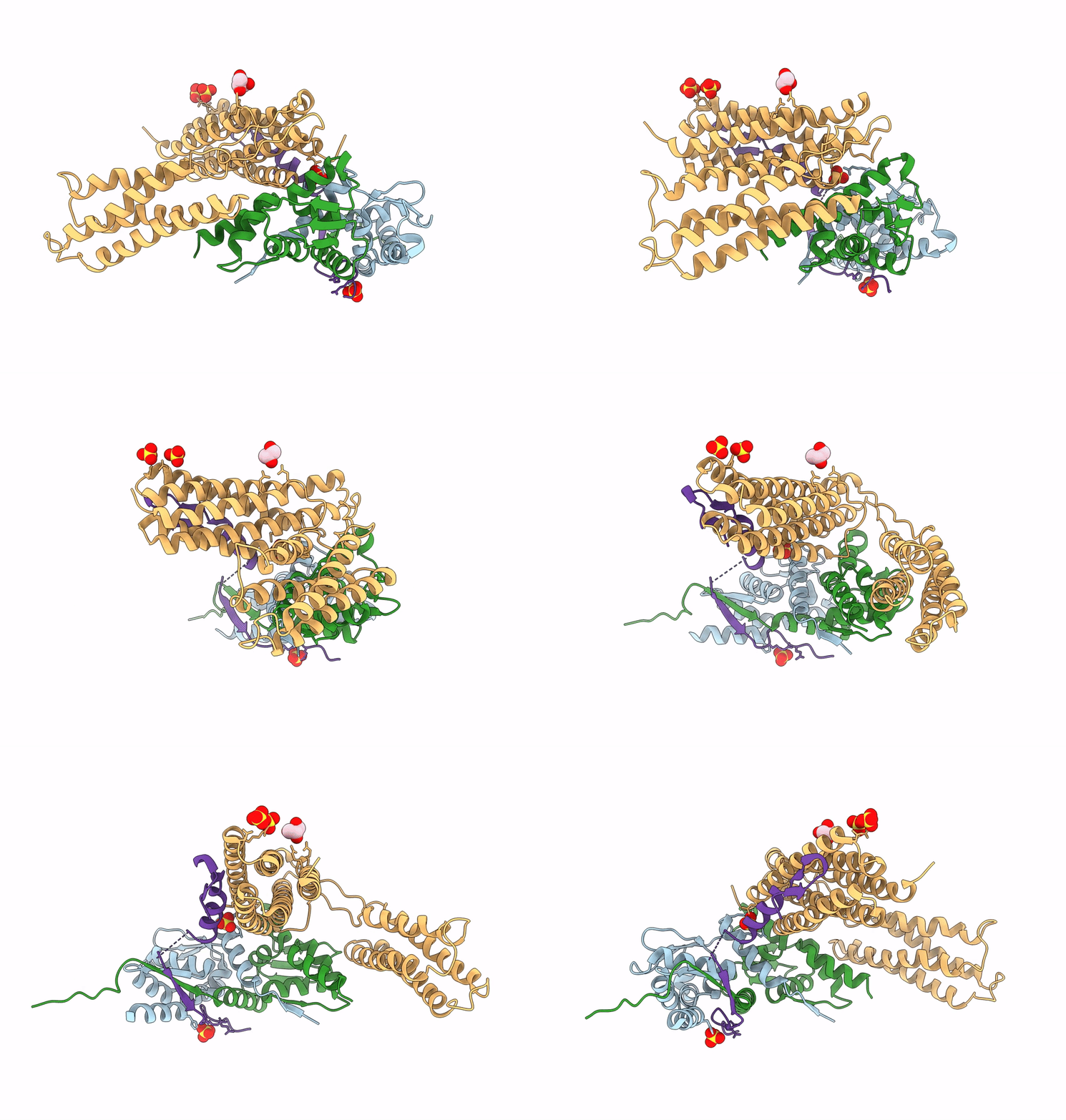
Crystal structure of the talin-KANK1 complex
Biological Source:
Source Organism:
Homo sapiens (Taxon ID: 9606)
Mus musculus (Taxon ID: 10090)
Method Details:
Experimental Method:
Resolution:
3.40 Å
R-Value Free:
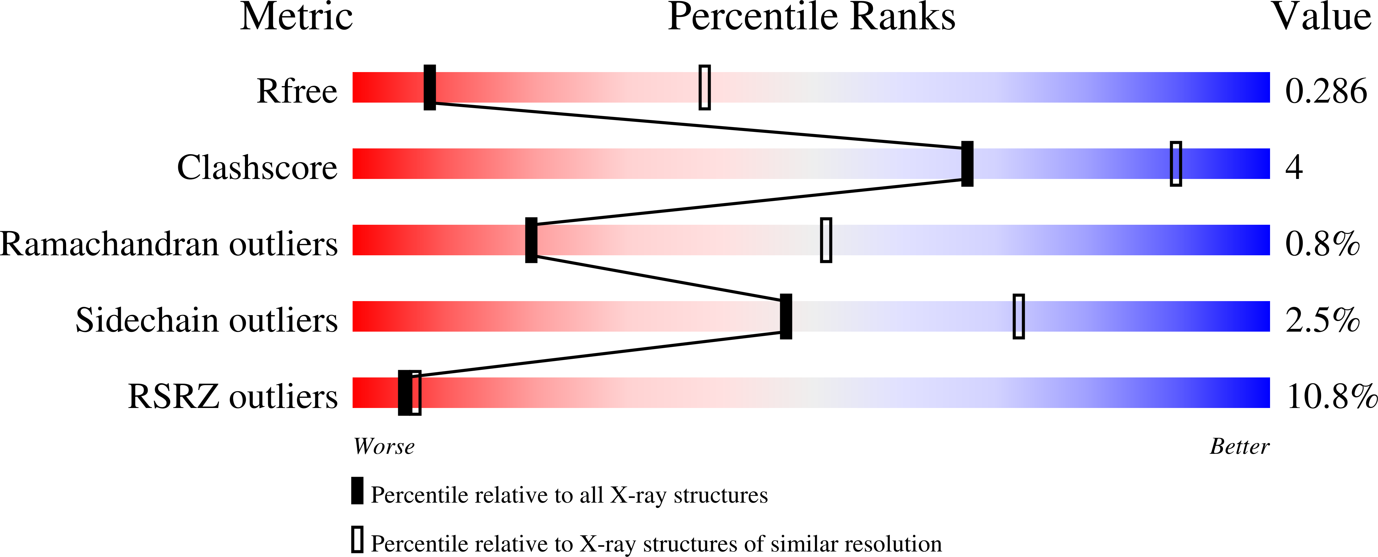
0.28
R-Value Work:
0.24
R-Value Observed:
0.24
Space Group:
H 3 2
Macromolecular Entities
Structures with similar UniProt ID
Protein Blast
Polymer Type:polypeptide(L)
Molecule:B-cell lymphoma 6 protein
Gene (Uniprot):BCL6
Chain IDs:A, B
Chain Length:137
Number of Molecules:2
Biological Source:Homo sapiens
Structures with similar UniProt ID
Protein Blast
Polymer Type:polypeptide(L)
Molecule:Talin-1
Gene (Uniprot):Tln1
Chain IDs:D (auth: C)
Chain Length:309
Number of Molecules:1
Biological Source:Mus musculus
Protein Blast
Polymer Type:polypeptide(L)
Molecule:KN-motif NCoR1 BBD fusion,Nuclear receptor corepressor 1
Gene (Uniprot):NCOR1
Chain IDs:C (auth: D)
Chain Length:51
Number of Molecules:1
Biological Source:Homo sapiens
Primary Citation
The structural basis of the talin-KANK1 interaction that coordinates the actin and microtubule cytoskeletons at focal adhesions.
Open Biology 13 230058 230058 (2023)
PMID: 37339751 DOI: 10.1098/rsob.230058

Abstact

Adhesion between cells and the extracellular matrix is mediated by heterodimeric (αβ) integrin receptors that are intracellularly linked to the contractile actomyosin machinery. One of the proteins that control this link is talin, which organizes cytosolic signalling proteins into discrete complexes on β-integrin tails referred to as focal adhesions (FAs). The adapter protein KANK1 binds to talin in the region of FAs known as the adhesion belt. Here, we adapted a non-covalent crystallographic chaperone to resolve the talin-KANK1 complex. This structure revealed that the talin binding KN region of KANK1 contains a novel motif where a β-hairpin stabilizes the α-helical region, explaining both its specific interaction with talin R7 and high affinity. Single point mutants in KANK1 identified from the structure abolished the interaction and enabled us to examine KANK1 enrichment in the adhesion belt. Strikingly, in cells expressing a constitutively active form of vinculin that keeps the FA structure intact even in the presence of myosin inhibitors, KANK1 localizes throughout the entire FA structure even when actomyosin tension is released. We propose a model whereby actomyosin forces on talin eliminate KANK1 from talin binding in the centre of FAs while retaining it at the adhesion periphery.

Legend

Protein

Chemical

Disease

Primary Citation of related structures