8AS3 image
Entry Detail
PDB ID:
8AS3
Title:
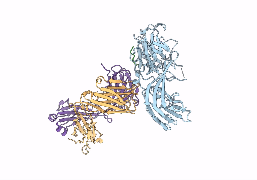
Structure of arrestin2 in complex with 6P CCR5 phosphopeptide and Fab30
Biological Source:
PDB Version:
Deposition Date:
2022-08-18
Release Date:
2023-06-07
Method Details:
Experimental Method:
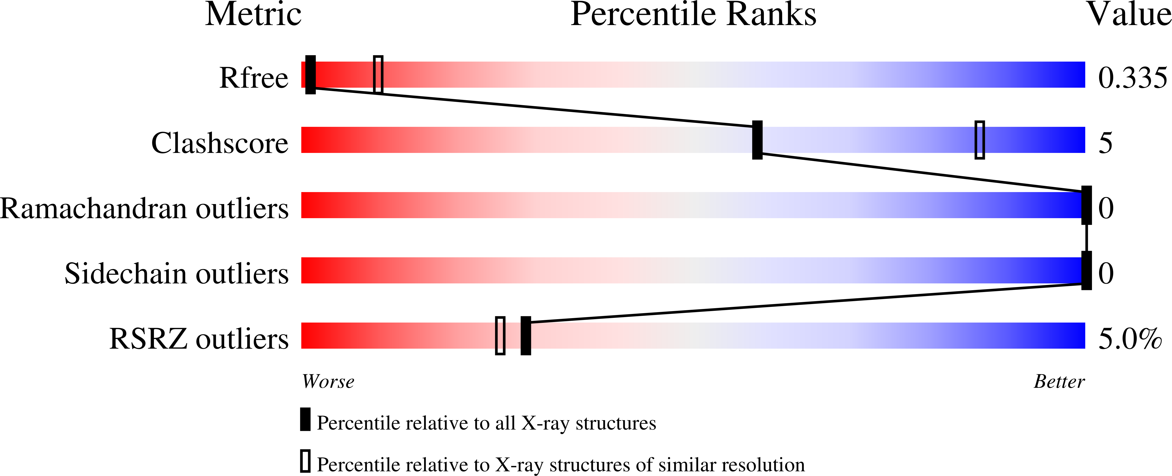
Resolution:
3.50 Å
R-Value Free:
0.33
R-Value Work:
0.28
R-Value Observed:
0.28
Space Group:
I 21 21 21
Macromolecular Entities
Polymer Type:polypeptide(L)
Description:Beta-arrestin-1
Chain IDs:A
Chain Length:359
Number of Molecules:1
Biological Source:Homo sapiens
Polymer Type:polypeptide(L)
Description:C-C chemokine receptor type 5
Chain IDs:B
Chain Length:21
Number of Molecules:1
Biological Source:Homo sapiens
Polymer Type:polypeptide(L)
Description:Fab30 heavy chain
Chain IDs:C (auth: H)
Chain Length:233
Number of Molecules:1
Biological Source:Phage display vector pTDisp
Polymer Type:polypeptide(L)
Description:Fab30 light chain
Chain IDs:D (auth: L)
Chain Length:220
Number of Molecules:1
Biological Source:Phage display vector pTDisp
Modified Residue
Compound ID Chain ID Parent Comp ID Details 2D Image
SEP B SER modified residue
TPO B THR modified residue
Ligand Molecules
Primary Citation
A key GPCR phosphorylation motif discovered in arrestin2⋅CCR5 phosphopeptide complexes.
Mol.Cell 83 2108 ? (2023)
PMID: 37244255 DOI: 10.1016/j.molcel.2023.05.002

Abstact

The two non-visual arrestins, arrestin2 and arrestin3, bind hundreds of GPCRs with different phosphorylation patterns, leading to distinct functional outcomes. Structural information on these interactions is available only for very few GPCRs. Here, we have characterized the interactions between the phosphorylated human CC chemokine receptor 5 (CCR5) and arrestin2. We identified several new CCR5 phosphorylation sites necessary for stable arrestin2 complex formation. Structures of arrestin2 in the apo form and complexes with CCR5 C-terminal phosphopeptides, together with NMR, biochemical, and functional assays, revealed three phosphoresidues in a pXpp motif that are essential for arrestin2 binding and activation. The identified motif appears responsible for robust arrestin2 recruitment in many other GPCRs. An analysis of receptor sequences and available structural and functional information provides hints on the molecular basis of arrestin2/arrestin3 isoform specificity. Our findings demonstrate how multi-site phosphorylation controls GPCR⋅arrestin interactions and provide a framework to probe the intricate details of arrestin signaling.

Legend

Protein

Chemical

Disease

Primary Citation of related structures