Deposition Date
2022-08-08
Release Date
2023-06-14
Last Version Date
2024-10-23
Entry Detail
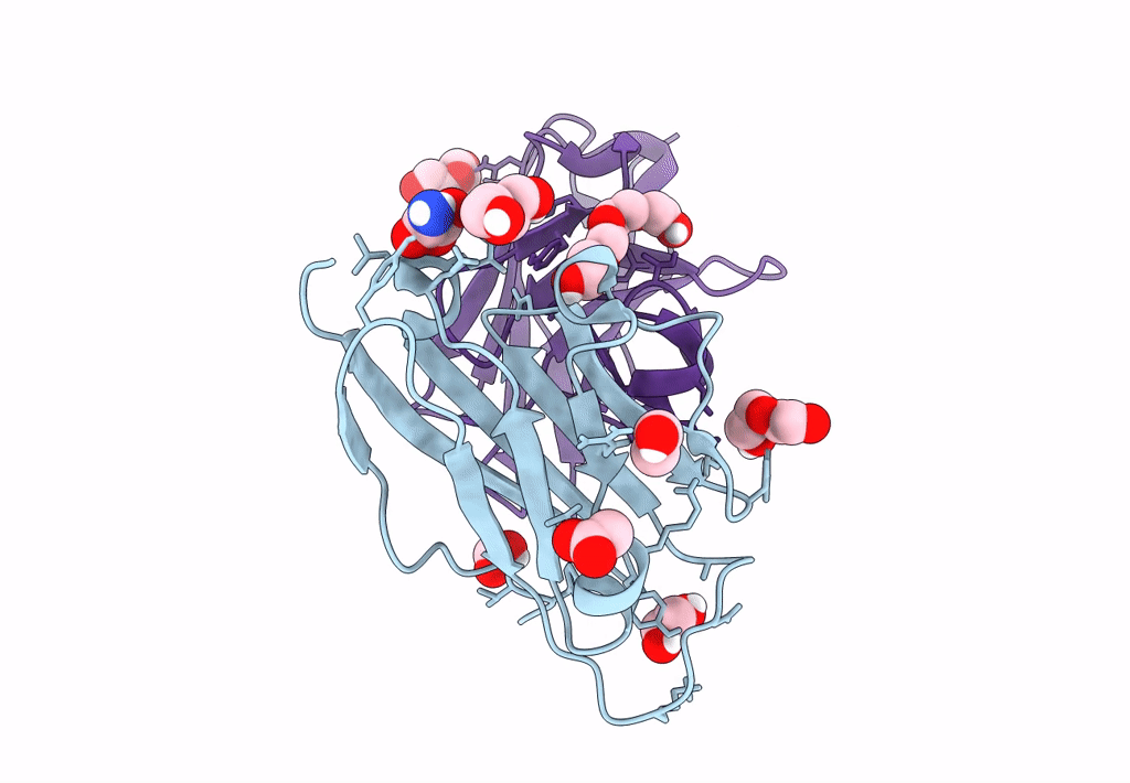
Biological Source:
Source Organism(s):
Homo sapiens (Taxon ID: 9606)
Lama glama (Taxon ID: 9844)
Lama glama (Taxon ID: 9844)
Expression System(s):
Method Details:
Experimental Method:
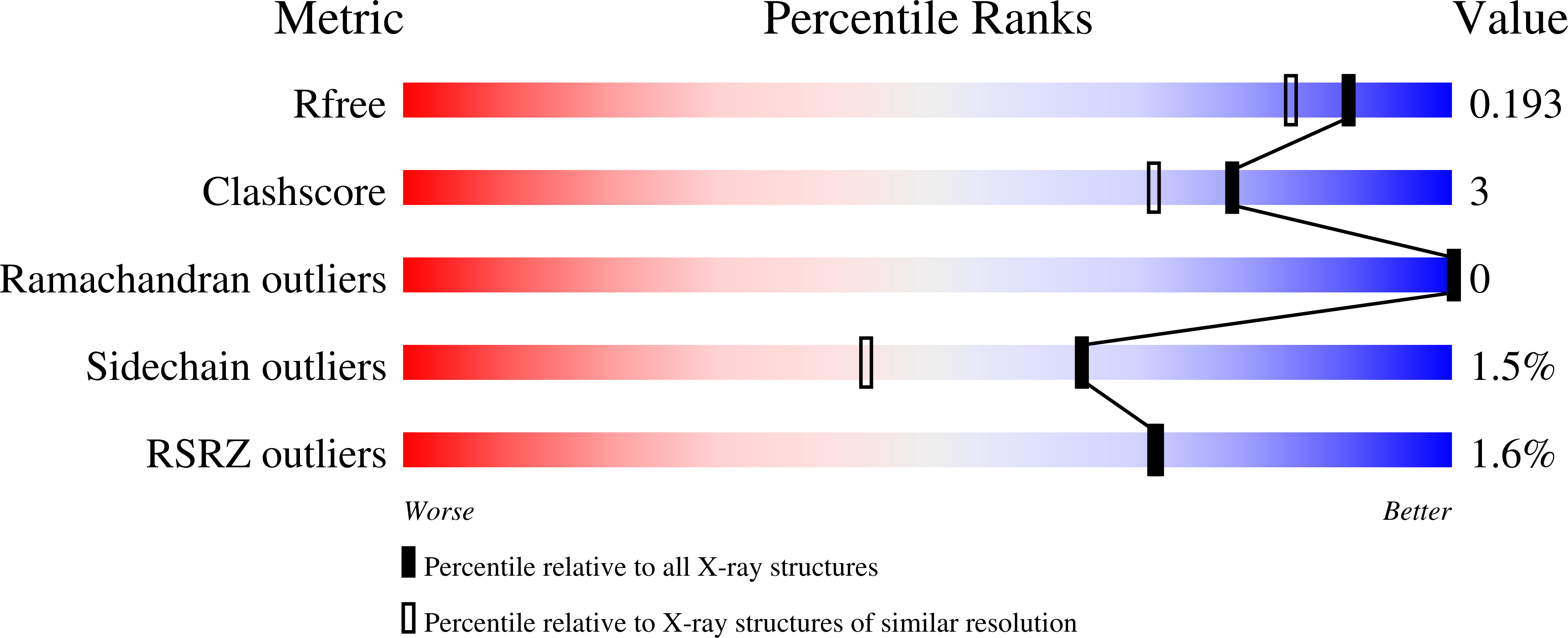
Resolution:
1.60 Å
R-Value Free:
0.18
R-Value Work:
0.16
Space Group:
I 41 2 2