8AMY image
Deposition Date 2022-08-04
Release Date 2023-08-23
Last Version Date 2024-10-16
Entry Detail
PDB ID:
8AMY
Keywords:
Title:
High-resolution crystal structure of the Mu8.1 conotoxin from Conus Mucronatus
Biological Source:
Source Organism:
Host Organism:
Method Details:
Experimental Method:
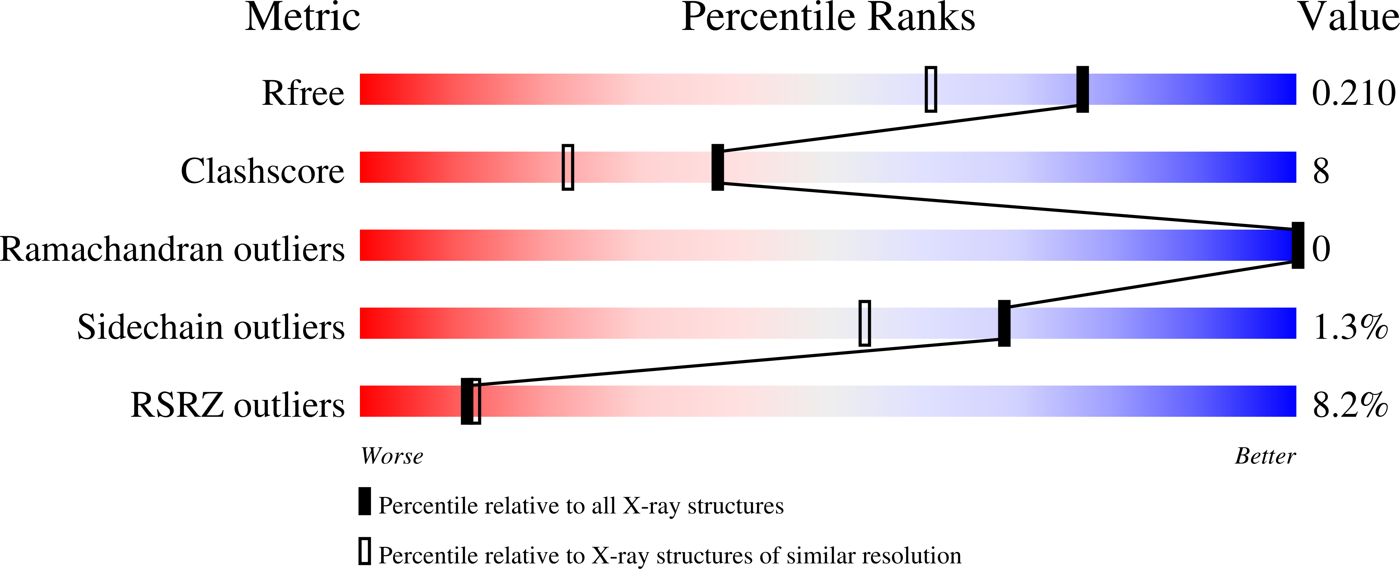
Resolution:
1.67 Å
R-Value Free:
0.20
R-Value Work:
0.16
R-Value Observed:
0.17
Space Group:
I 41 2 2
Macromolecular Entities
Polymer Type:polypeptide(L)
Molecule:Mu8.1 conotoxin
Chain IDs:A
Chain Length:89
Number of Molecules:1
Biological Source:Conus mucronatus
Primary Citation
High-resolution crystal structure of the Mu8.1 conotoxin from Conus mucronatus.
Acta Crystallogr.,Sect.F 79 240 246 (2023)
PMID: 37642664 DOI: 10.1107/S2053230X23007070

Abstact

Marine cone snails produce a wealth of peptide toxins (conotoxins) that bind their molecular targets with high selectivity and potency. Therefore, conotoxins constitute valuable biomolecular tools with a variety of biomedical purposes. The Mu8.1 conotoxin from Conus mucronatus is the founding member of the newly identified saposin-like conotoxin class of conotoxins and has been shown to target Cav2.3, a voltage-gated calcium channel. Two crystal structures have recently been determined of Mu8.1 at 2.3 and 2.1 Å resolution. Here, a high-resolution crystal structure of Mu8.1 was determined at 1.67 Å resolution in the high-symmetry space group I4122. The asymmetric unit contained one molecule, with a symmetry-related molecule generating a dimer equivalent to that observed in the two previously determined structures. The high resolution allows a detailed atomic analysis of a water-filled cavity buried at the dimer interface, revealing a tightly coordinated network of waters that shield a lysine residue (Lys55) with a predicted unusually low side-chain pKa value. These findings are discussed in terms of a potential functional role of Lys55 in target interaction.

Legend

Protein

Chemical

Disease

Primary Citation of related structures