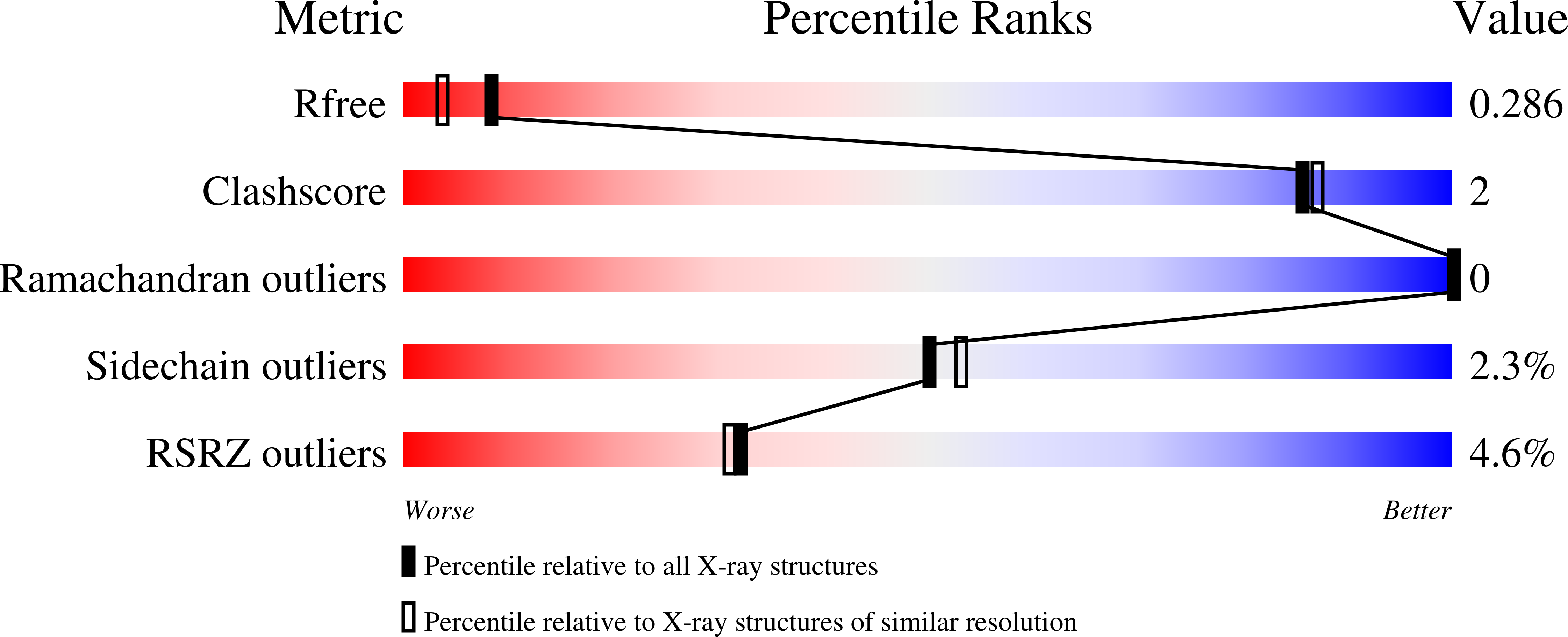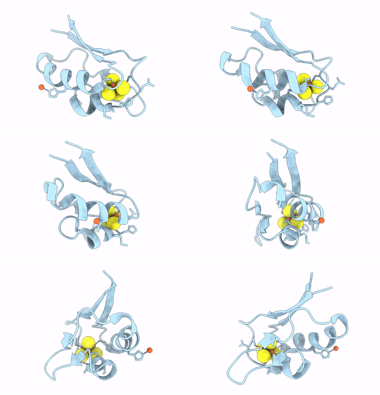
8AMP image
Deposition Date 2022-08-03
Release Date 2023-02-15
Last Version Date 2024-02-07
Entry Detail
PDB ID:
8AMP
Title:
Crystal structure of M.tuberculosis ferredoxin Fdx
Biological Source:
Source Organism:
Host Organism:
Method Details:
Experimental Method:
Resolution:
2.00 Å
R-Value Free:
0.29
R-Value Work:
0.25
R-Value Observed:
0.25
Space Group:
H 3 2
Macromolecular Entities
Polymer Type:polypeptide(L)
Molecule:Possible ferredoxin
Gene (Uniprot):fdx
Chain IDs:A
Chain Length:74
Number of Molecules:1
Biological Source:Mycobacterium tuberculosis
Primary Citation
Structural insights into 3Fe-4S ferredoxins diversity in M. tuberculosis highlighted by a first redox complex with P450.
Front Mol Biosci 9 1100032 1100032 (2022)
PMID: 36699703 DOI: 10.3389/fmolb.2022.1100032

Abstact

Ferredoxins are small iron-sulfur proteins and key players in essential metabolic pathways. Among all types, 3Fe-4S ferredoxins are less studied mostly due to anaerobic requirements. Their complexes with cytochrome P450 redox partners have not been structurally characterized. In the present work, we solved the structures of both 3Fe-4S ferredoxins from M. tuberculosis-Fdx alone and the fusion FdxE-CYP143. Our SPR analysis demonstrated a high-affinity binding of FdxE to CYP143. According to SAXS data, the same complex is present in solution. The structure reveals extended multipoint interactions and the shape/charge complementarity of redox partners. Furthermore, FdxE binding induced conformational changes in CYP143 as evident from the solved CYP143 structure alone. The comparison of FdxE-CYP143 and modeled Fdx-CYP51 complexes further revealed the specificity of ferredoxins. Our results illuminate the diversity of electron transfer complexes for the production of different secondary metabolites.

Legend

Protein

Chemical

Disease

Primary Citation of related structures