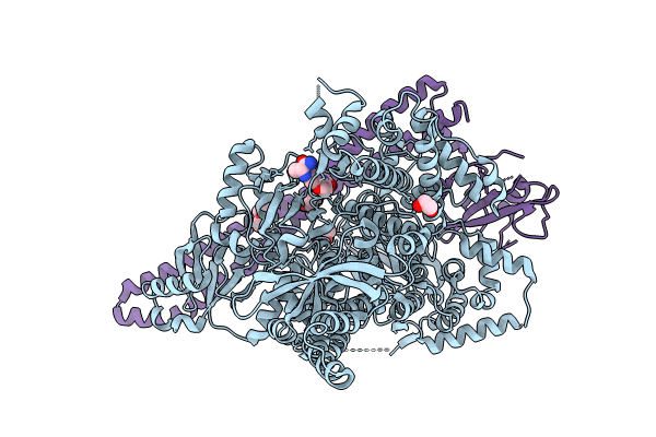
8AM0 image
Entry Detail
PDB ID:
8AM0
Keywords:
Title:
Crystal structure of human T1061E PI3Kalpha in complex with its regulatory subunit and the inhibitor GDC-0077 (Inavolisib)
Biological Source:
Source Organism:
Host Organism:
PDB Version:
Deposition Date:
2022-08-02
Release Date:
2023-12-13
Method Details:
Experimental Method:
Resolution:
2.82 Å
R-Value Free:
0.30
R-Value Work:
0.28
R-Value Observed:
0.28
Space Group:
P 21 21 21
Macromolecular Entities
Polymer Type:polypeptide(L)
Description:Phosphatidylinositol 4,5-bisphosphate 3-kinase catalytic subunit alpha isoform
Mutations:T1061E
Chain IDs:A
Chain Length:1068
Number of Molecules:1
Biological Source:Homo sapiens
Polymer Type:polypeptide(L)
Description:Isoform 3 of Phosphatidylinositol 3-kinase regulatory subunit alpha
Chain IDs:B
Chain Length:293
Number of Molecules:1
Biological Source:Homo sapiens
Primary Citation

Abstact

The phosphoinositide 3-kinase p110α is an essential mediator of insulin signaling and glucose homeostasis. We interrogated the human serine, threonine, and tyrosine kinome to search for novel regulators of p110α and found that the Hippo kinases phosphorylate p110α at T1061, which inhibits its activity. This inhibitory state corresponds to a conformational change of a membrane-binding domain on p110α, which impairs its ability to engage membranes. In human primary hepatocytes, cancer cell lines, and rodent tissues, activation of the Hippo kinases MST1/2 using forskolin or epinephrine is associated with phosphorylation of T1061 and inhibition of p110α, impairment of downstream insulin signaling, and suppression of glycolysis and glycogen synthesis. These changes are abrogated when MST1/2 are genetically deleted or inhibited with small molecules or if the T1061 is mutated to alanine. Our study defines an inhibitory pathway of PI3K signaling and a link between epinephrine and insulin signaling.

Legend

Protein

Chemical

Disease

Primary Citation of related structures