8AKO image
Deposition Date 2022-07-30
Release Date 2022-12-14
Last Version Date 2024-01-31
Entry Detail
PDB ID:
8AKO
Keywords:
Title:
Structure of EspB-EspK complex: the non-identical twin of the PE-PPE-EspG secretion mechanism.
Biological Source:
Source Organism:
Host Organism:
Method Details:
Experimental Method:
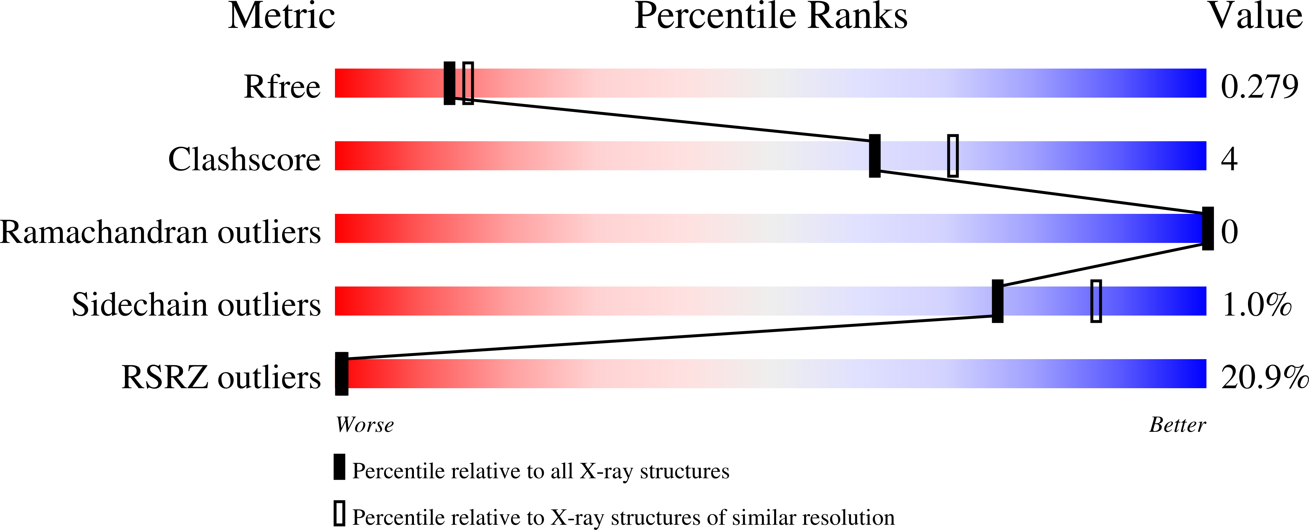
Resolution:
2.29 Å
R-Value Free:
0.27
R-Value Work:
0.23
Space Group:
P 61 2 2
Macromolecular Entities
Structures with similar UniProt ID
Protein Blast
Polymer Type:polypeptide(L)
Molecule:ESX-1 secretion-associated protein EspB
Gene (Uniprot):espB
Chain IDs:A
Chain Length:300
Number of Molecules:1
Biological Source:Mycobacterium tuberculosis
Structures with similar UniProt ID
Protein Blast
Polymer Type:polypeptide(L)
Molecule:ESX-1 secretion-associated protein EspK
Gene (Uniprot):espK
Chain IDs:B
Chain Length:246
Number of Molecules:1
Biological Source:Mycobacterium tuberculosis
Primary Citation
The crystal structure of the EspB-EspK virulence factor-chaperone complex suggests an additional type VII secretion mechanism in Mycobacterium tuberculosis.
J.Biol.Chem. 299 102761 102761 (2022)
PMID: 36463964 DOI: 10.1016/j.jbc.2022.102761

Abstact

Pathogenic species from the Mycobacterium genus are responsible for a number of adverse health conditions in humans and animals that threaten health security and the economy worldwide. Mycobacteria have up to five specialized secretion systems (ESX-1 to ESX-5) that transport virulence factors across their complex cell envelope to facilitate manipulation of their environment. In pathogenic species, these virulence factors influence the immune system's response and are responsible for membrane disruption and contributing to cell death. While structural details of these secretion systems have been recently described, gaps still remain in the structural understanding of the secretion mechanisms of most substrates. Here, we describe the crystal structure of Mycobacterium tuberculosis ESX-1 secretion-associated substrate EspB bound to its chaperone EspK. We found that EspB interacts with the C-terminal domain of EspK through its helical tip. Furthermore, cryogenic electron microscopy, size exclusion chromatography analysis, and small-angle X-ray scattering experiments show that EspK keeps EspB in its secretion-competent monomeric form and prevents its oligomerization. The structure presented in this study suggests an additional secretion mechanism in ESX-1, analogous to the chaperoning of proline-glutamate (PE)-proline-proline-glutamate (PPE) proteins by EspG, where EspK facilitates the secretion of EspB in Mycobacterium species.

Legend

Protein

Chemical

Disease

Primary Citation of related structures