8ADN image
Deposition Date 2022-07-08
Release Date 2022-11-23
Last Version Date 2024-10-16
Entry Detail
PDB ID:
8ADN
Keywords:
Title:
Vairimorpha necatrix 20S proteasome from spores
Biological Source:
Source Organism:
Host Organism:
Method Details:
Experimental Method:
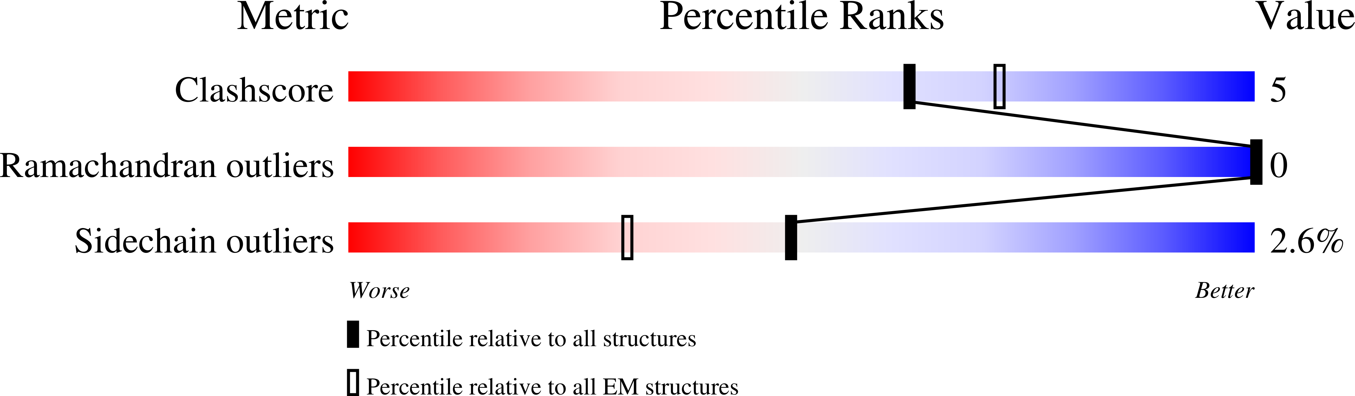
Resolution:
2.77 Å
Aggregation State:
PARTICLE
Reconstruction Method:
SINGLE PARTICLE
Macromolecular Entities
Polymer Type:polypeptide(L)
Molecule:Proteasome Inhibitor 31-Like
Chain IDs:A (auth: 3), B (auth: 4)
Chain Length:147
Number of Molecules:2
Biological Source:Vairimorpha necatrix
Polymer Type:polypeptide(L)
Molecule:Proteasome subunit alpha type-2
Chain IDs:C (auth: A), Q (auth: O)
Chain Length:212
Number of Molecules:2
Biological Source:Vairimorpha necatrix
Polymer Type:polypeptide(L)
Molecule:Proteasome subunit alpha type-3
Chain IDs:D (auth: B), R (auth: P)
Chain Length:231
Number of Molecules:2
Biological Source:Vairimorpha necatrix
Polymer Type:polypeptide(L)
Molecule:Proteasome subunit alpha type-4
Chain IDs:E (auth: C), S (auth: Q)
Chain Length:225
Number of Molecules:2
Biological Source:Vairimorpha necatrix
Polymer Type:polypeptide(L)
Molecule:Proteasome subunit alpha type-5
Chain IDs:F (auth: D), T (auth: R)
Chain Length:234
Number of Molecules:2
Biological Source:Vairimorpha necatrix
Polymer Type:polypeptide(L)
Molecule:Proteasome subunit alpha type-6
Chain IDs:G (auth: E), U (auth: S)
Chain Length:230
Number of Molecules:2
Biological Source:Vairimorpha necatrix
Polymer Type:polypeptide(L)
Molecule:Proteasome subunit alpha type-7
Chain IDs:H (auth: F), V (auth: T)
Chain Length:243
Number of Molecules:2
Biological Source:Vairimorpha necatrix
Polymer Type:polypeptide(L)
Molecule:Proteasome subunit alpha type-1
Chain IDs:I (auth: G), W (auth: U)
Chain Length:234
Number of Molecules:2
Biological Source:Vairimorpha necatrix
Polymer Type:polypeptide(L)
Molecule:Proteasome subunit beta type-2
Chain IDs:J (auth: H), X (auth: V)
Chain Length:227
Number of Molecules:2
Biological Source:Vairimorpha necatrix
Polymer Type:polypeptide(L)
Molecule:Proteasome subunit beta type-3
Chain IDs:K (auth: I), Y (auth: W)
Chain Length:205
Number of Molecules:2
Biological Source:Vairimorpha necatrix
Polymer Type:polypeptide(L)
Molecule:Proteasome subunit beta type-4
Chain IDs:L (auth: J), Z (auth: X)
Chain Length:193
Number of Molecules:2
Biological Source:Vairimorpha necatrix
Polymer Type:polypeptide(L)
Molecule:Proteasome subunit beta type-5
Chain IDs:M (auth: K), AA (auth: Y)
Chain Length:228
Number of Molecules:2
Biological Source:Vairimorpha necatrix
Polymer Type:polypeptide(L)
Molecule:Proteasome subunit beta type-6
Chain IDs:N (auth: L), BA (auth: Z)
Chain Length:297
Number of Molecules:2
Biological Source:Vairimorpha necatrix
Polymer Type:polypeptide(L)
Molecule:Proteasome subunit beta type-7
Chain IDs:O (auth: M), CA (auth: a)
Chain Length:212
Number of Molecules:2
Biological Source:Vairimorpha necatrix
Polymer Type:polypeptide(L)
Molecule:Proteasome subunit beta type-1
Chain IDs:P (auth: N), DA (auth: b)
Chain Length:231
Number of Molecules:2
Biological Source:Vairimorpha necatrix
Ligand Molecules
Primary Citation
Structure of the reduced microsporidian proteasome bound by PI31-like peptides in dormant spores.
Nat Commun 13 6962 6962 (2022)
PMID: 36379934 DOI: 10.1038/s41467-022-34691-x

Abstact

Proteasomes play an essential role in the life cycle of intracellular pathogens with extracellular stages by ensuring proteostasis in environments with limited resources. In microsporidia, divergent parasites with extraordinarily streamlined genomes, the proteasome complexity and structure are unknown, which limits our understanding of how these unique pathogens adapt and compact essential eukaryotic complexes. We present cryo-electron microscopy structures of the microsporidian 20S and 26S proteasome isolated from dormant or germinated Vairimorpha necatrix spores. The discovery of PI31-like peptides, known to inhibit proteasome activity, bound simultaneously to all six active sites within the central cavity of the dormant spore proteasome, suggests reduced activity in the environmental stage. In contrast, the absence of the PI31-like peptides and the existence of 26S particles post-germination in the presence of ATP indicates that proteasomes are reactivated in nutrient-rich conditions. Structural and phylogenetic analyses reveal that microsporidian proteasomes have undergone extensive reductive evolution, lost at least two regulatory proteins, and compacted nearly every subunit. The highly derived structure of the microsporidian proteasome, and the minimized version of PI31 presented here, reinforce the feasibility of the development of specific inhibitors and provide insight into the unique evolution and biology of these medically and economically important pathogens.

Legend

Protein

Chemical

Disease

Primary Citation of related structures
Feedback Form
Name
Email
Institute
Feedback