Deposition Date
2022-06-30
Release Date
2022-10-19
Last Version Date
2024-07-24
Entry Detail
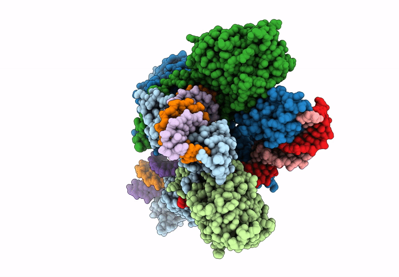
PDB ID:
8AA5
Keywords:
Title:
Cryo-EM structure of the strand transfer complex of the TnsB transposase (type V-K CRISPR-associated transposon)
Biological Source:
Source Organism(s):
Scytonema hofmannii (Taxon ID: 34078)
Expression System(s):
Method Details:
Experimental Method:
Resolution:
2.46 Å
Aggregation State:
PARTICLE
Reconstruction Method:
SINGLE PARTICLE