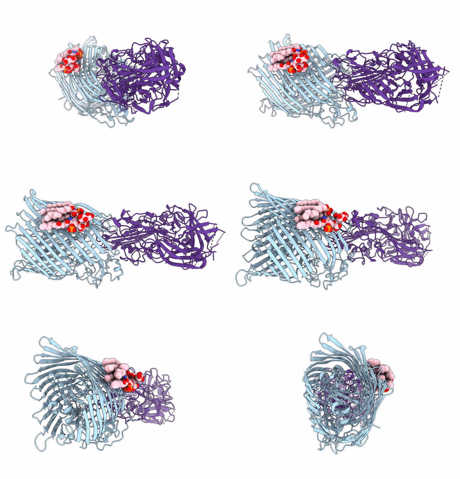
8A8C image
Deposition Date 2022-06-22
Release Date 2022-10-05
Last Version Date 2024-11-20
Entry Detail
PDB ID:
8A8C
Title:
T5 phage receptor-binding protein pb5 bound to ferrichrome transporter FhuA
Biological Source:
Source Organism:
Host Organism:
Method Details:
Experimental Method:
Resolution:
3.10 Å
Aggregation State:
PARTICLE
Reconstruction Method:
SINGLE PARTICLE
Macromolecular Entities
Structures with similar UniProt ID
Protein Blast
Polymer Type:polypeptide(L)
Molecule:Ferrichrome outer membrane transporter/phage receptor
Gene (Uniprot):fhuA
Chain IDs:A
Chain Length:714
Number of Molecules:1
Biological Source:Escherichia coli K-12
Structures with similar UniProt ID
Protein Blast
Polymer Type:polypeptide(L)
Molecule:Receptor-binding protein pb5
Gene (Uniprot):oad
Chain IDs:B
Chain Length:646
Number of Molecules:1
Biological Source:Escherichia phage T5
Ligand Molecules
Primary Citation
Structural basis for host recognition and superinfection exclusion by bacteriophage T5.
Proc.Natl.Acad.Sci.USA 119 e2211672119 e2211672119 (2022)
PMID: 36215462 DOI: 10.1073/pnas.2211672119

Abstact

A key but poorly understood stage of the bacteriophage life cycle is the binding of phage receptor-binding proteins (RBPs) to receptors on the host cell surface, leading to injection of the phage genome and, for lytic phages, host cell lysis. To prevent secondary infection by the same or a closely related phage and nonproductive phage adsorption to lysed cell fragments, superinfection exclusion (SE) proteins can prevent the binding of RBPs via modulation of the host receptor structure in ways that are also unclear. Here, we present the cryogenic electron microscopy (cryo-EM) structure of the phage T5 outer membrane (OM) receptor FhuA in complex with the T5 RBP pb5, and the crystal structure of FhuA complexed to the OM SE lipoprotein Llp. Pb5 inserts four loops deeply into the extracellular lumen of FhuA and contacts the plug but does not cause any conformational changes in the receptor, supporting the view that DNA translocation does not occur through the lumen of OM channels. The FhuA-Llp structure reveals that Llp is periplasmic and binds to a nonnative conformation of the plug of FhuA, causing the inward folding of two extracellular loops via "reverse" allostery. The inward-folded loops of FhuA overlap with the pb5 binding site, explaining how Llp binding to FhuA abolishes further infection of Escherichia coli by phage T5 and suggesting a mechanism for SE via the jamming of TonB-dependent transporters by small phage lipoproteins.

Legend

Protein

Chemical

Disease

Primary Citation of related structures