8A39 image
Deposition Date 2022-06-07
Release Date 2023-07-05
Last Version Date 2024-01-17
Entry Detail
PDB ID:
8A39
Keywords:
Title:
Crystal Structure of PaaX from Escherichia coli W
Biological Source:
Source Organism:
Host Organism:
Method Details:
Experimental Method:
Resolution:
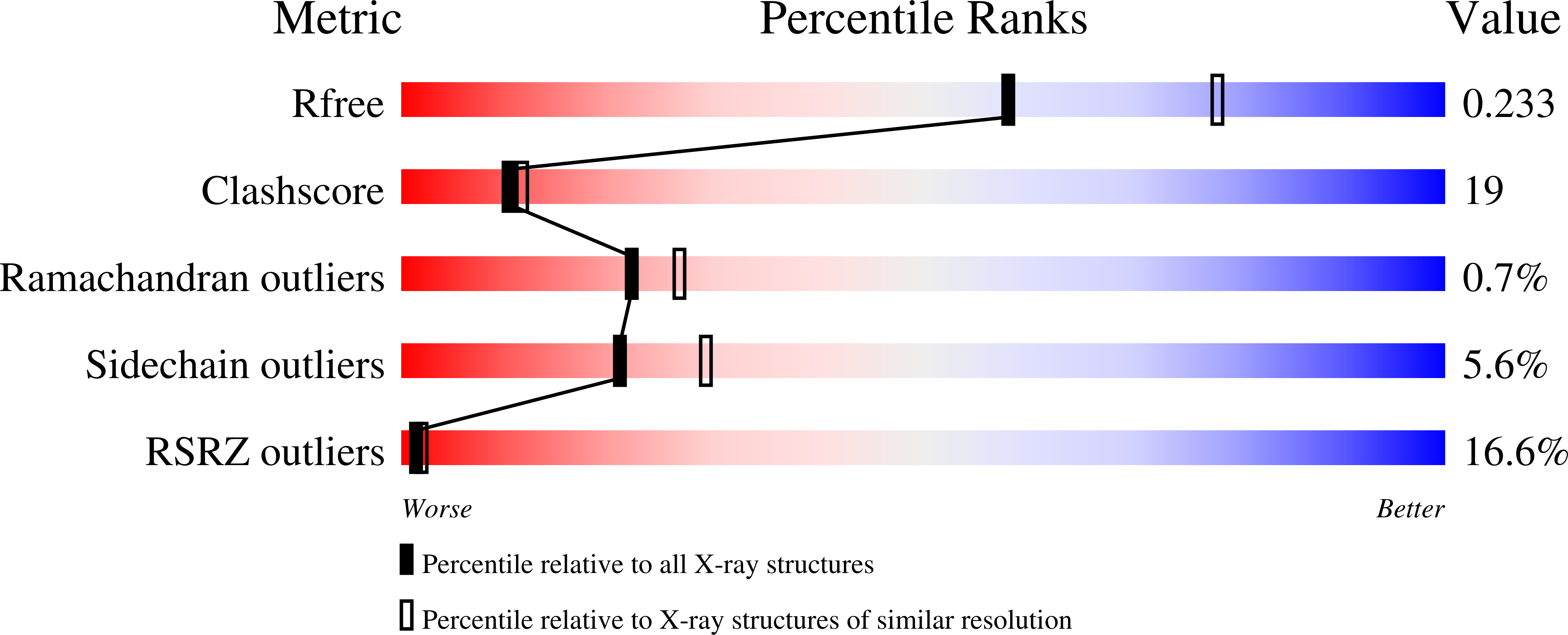
2.30 Å
R-Value Free:
0.24
R-Value Work:
0.19
R-Value Observed:
0.19
Space Group:
C 1 2 1
Macromolecular Entities
Structures with similar UniProt ID
Protein Blast
Polymer Type:polypeptide(L)
Molecule:DNA-binding transcriptional repressor of phenylacetic acid degradation, aryl-CoA responsive
Gene (Uniprot):paaX
Chain IDs:A (auth: AP1), B (auth: BP1), C (auth: CP1)
Chain Length:316
Number of Molecules:3
Biological Source:Escherichia coli W
Primary Citation
Structural characterization of PaaX, the main repressor of the phenylacetate degradation pathway in Escherichia coli W: A novel fold of transcription regulator proteins.
Int.J.Biol.Macromol. 254 127935 127935 (2024)
PMID: 37949283 DOI: 10.1016/j.ijbiomac.2023.127935

Abstact

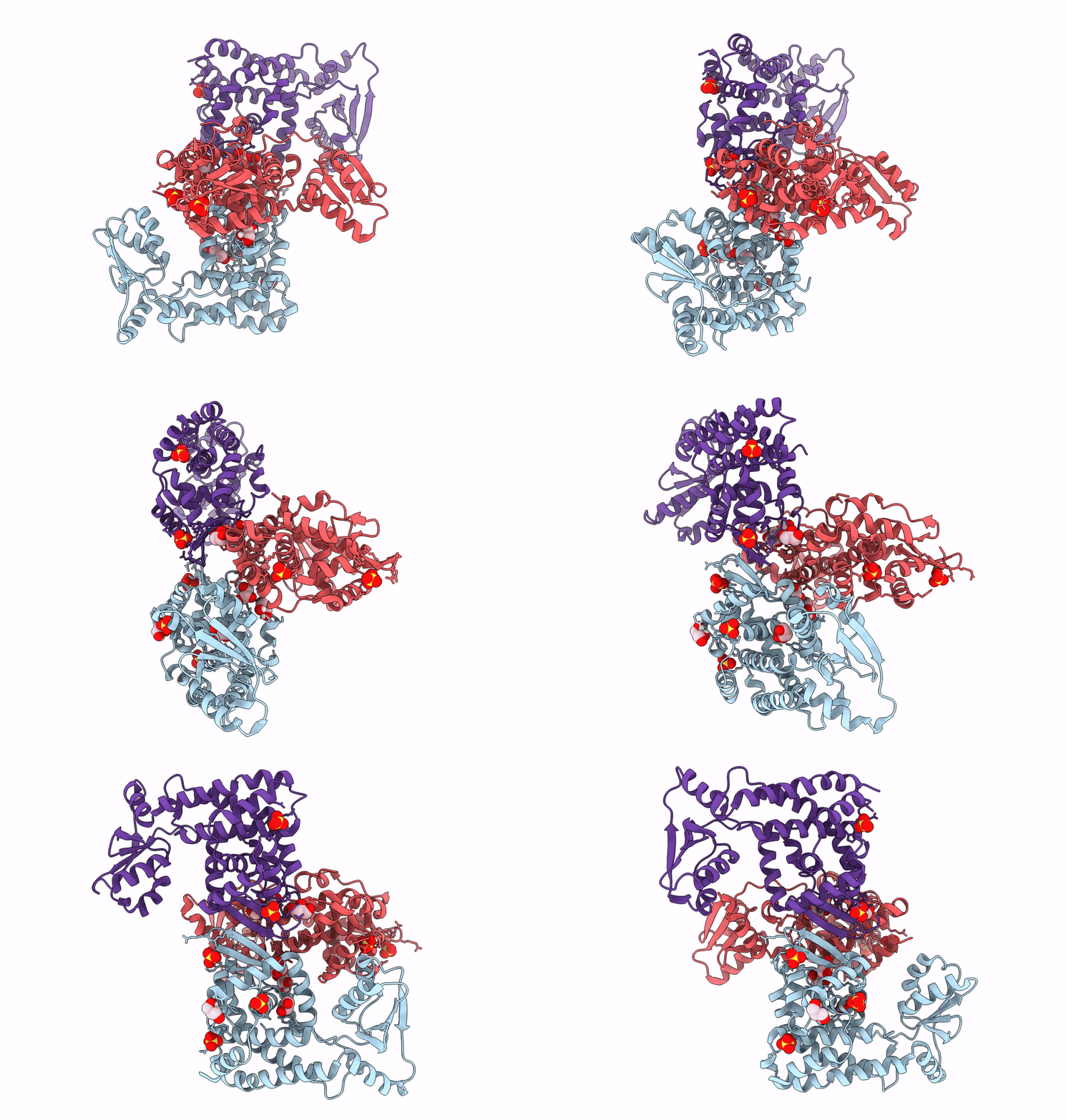
PaaX is a transcriptional repressor of the phenylacetic acid (PAA) catabolic pathway, a central route for bacterial aerobic degradation of aromatic compounds. Induction of the route is achieved through the release of PaaX from its promoter sequences by the first compound of the pathway, phenylacetyl-coenzyme A (PA-CoA). We report the crystal structure of PaaX from Escherichia coli W. PaaX displays a novel type of fold for transcription regulators, showing a dimeric conformation where the monomers present a three-domain structure: an N-terminal winged helix-turn-helix domain, a dimerization domain similar to the Cas2 protein and a C-terminal domain without structural homologs. The domains are separated by a crevice amenable to harbour a PA-CoA molecule. The biophysical characterization of the protein in solution confirmed several hints predicted from the structure, i.e. its dimeric conformation, a modest importance of cysteines and a high dependence of solubility and thermostability on ionic strength. At a moderately acidic pH, the protein formed a stable folding intermediate with remaining α-helical structure, a disrupted tertiary structure and exposed hydrophobic patches. Our results provide valuable information to understand the stability and mechanism of PaaX and pave the way for further analysis of other regulators with similar structural configurations.

Legend

Protein

Chemical

Disease

Primary Citation of related structures