7awk image
Deposition Date 2020-11-08
Release Date 2021-11-24
Last Version Date 2024-06-19
Entry Detail
PDB ID:
7AWK
Keywords:
Title:
Crystal structure of the HigB1 toxin mutant K95A from Mycobacterium tuberculosis (Rv1955)
Biological Source:
Method Details:
Experimental Method:
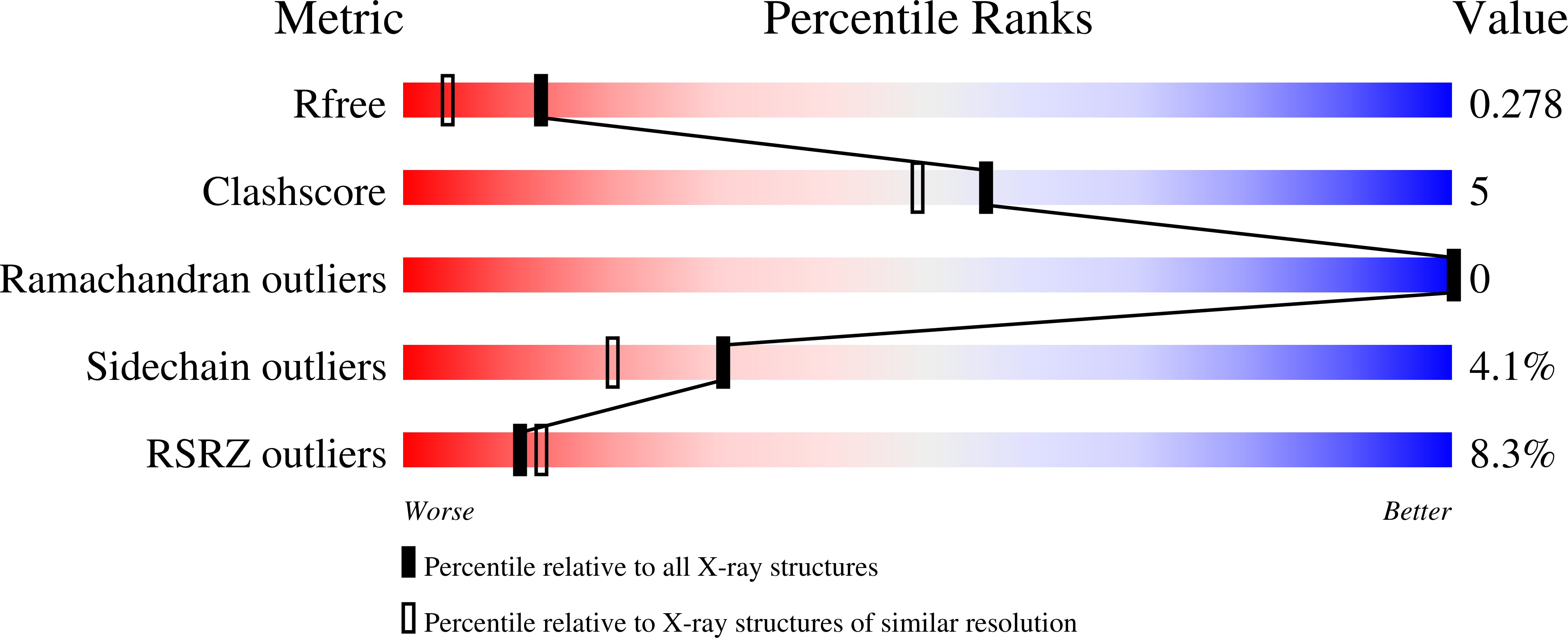
Resolution:
1.91 Å
R-Value Free:
0.27
R-Value Work:
0.21
Space Group:
P 21 21 2
Macromolecular Entities
Polymer Type:polypeptide(L)
Molecule:Probable endoribonuclease HigB1
Gene (Uniprot):higB1
Mutations:K95A
Chain IDs:A
Chain Length:130
Number of Molecules:1
Biological Source:Mycobacterium tuberculosis (strain ATCC 25618 / H37Rv)
Primary Citation
Substrate recognition and cryo-EM structure of the ribosome-bound TAC toxin of Mycobacterium tuberculosis.
Nat Commun 13 2641 2641 (2022)
PMID: 35552387 DOI: 10.1038/s41467-022-30373-w

Abstact

Toxins of toxin-antitoxin systems use diverse mechanisms to control bacterial growth. Here, we focus on the deleterious toxin of the atypical tripartite toxin-antitoxin-chaperone (TAC) system of Mycobacterium tuberculosis, whose inhibition requires the concerted action of the antitoxin and its dedicated SecB-like chaperone. We show that the TAC toxin is a bona fide ribonuclease and identify exact cleavage sites in mRNA targets on a transcriptome-wide scale in vivo. mRNA cleavage by the toxin occurs after the second nucleotide of the ribosomal A-site codon during translation, with a strong preference for CCA codons in vivo. Finally, we report the cryo-EM structure of the ribosome-bound TAC toxin in the presence of native M. tuberculosis cspA mRNA, revealing the specific mechanism by which the TAC toxin interacts with the ribosome and the tRNA in the P-site to cleave its mRNA target.

Legend

Protein

Chemical

Disease

Primary Citation of related structures