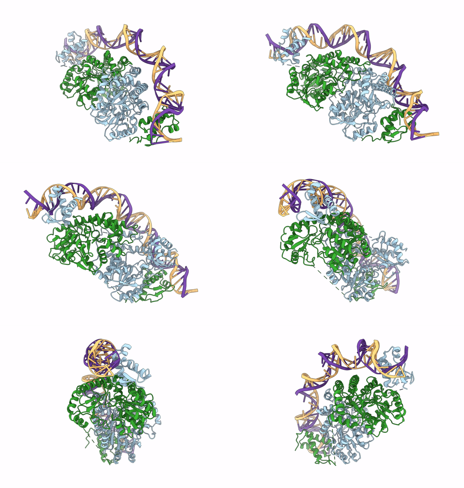
7ZPA image
Entry Detail
PDB ID:
7ZPA
EMDB ID:
Title:
Cryo-EM structure of holo-PdxR from Bacillus clausii bound to its target DNA in the closed conformation, C1 symmetry
Biological Source:
PDB Version:
Deposition Date:
2022-04-27
Release Date:
2023-07-05
Method Details:
Experimental Method:
Resolution:
3.90 Å
Aggregation State:
PARTICLE
Reconstruction Method:
SINGLE PARTICLE
Macromolecular Entities
Polymer Type:polypeptide(L)
Description:PLP-dependent aminotransferase family protein
Chain IDs:A, B
Chain Length:478
Number of Molecules:2
Biological Source:Alkalihalobacillus clausii
Polymer Type:polydeoxyribonucleotide
Description:DNA (48-MER)
Chain IDs:C
Chain Length:48
Number of Molecules:1
Biological Source:Alkalihalobacillus clausii
Polymer Type:polydeoxyribonucleotide
Description:DNA (48-MER)
Chain IDs:D
Chain Length:48
Number of Molecules:1
Biological Source:Alkalihalobacillus clausii
Modified Residue
Compound ID Chain ID Parent Comp ID Details 2D Image
LLP A LYS modified residue
Ligand Molecules
Primary Citation
Structural insights into the DNA recognition mechanism by the bacterial transcription factor PdxR.
Nucleic Acids Res. 51 8237 8254 (2023)
PMID: 37378428 DOI: 10.1093/nar/gkad552

Abstact

Specificity in protein-DNA recognition arises from the synergy of several factors that stem from the structural and chemical signatures encoded within the targeted DNA molecule. Here, we deciphered the nature of the interactions driving DNA recognition and binding by the bacterial transcription factor PdxR, a member of the MocR family responsible for the regulation of pyridoxal 5'-phosphate (PLP) biosynthesis. Single particle cryo-EM performed on the PLP-PdxR bound to its target DNA enabled the isolation of three conformers of the complex, which may be considered as snapshots of the binding process. Moreover, the resolution of an apo-PdxR crystallographic structure provided a detailed description of the transition of the effector domain to the holo-PdxR form triggered by the binding of the PLP effector molecule. Binding analyses of mutated DNA sequences using both wild type and PdxR variants revealed a central role of electrostatic interactions and of the intrinsic asymmetric bending of the DNA in allosterically guiding the holo-PdxR-DNA recognition process, from the first encounter through the fully bound state. Our results detail the structure and dynamics of the PdxR-DNA complex, clarifying the mechanism governing the DNA-binding mode of the holo-PdxR and the regulation features of the MocR family of transcription factors.

Legend

Protein

Chemical

Disease

Primary Citation of related structures