7ZP0 image
Deposition Date 2022-04-26
Release Date 2022-08-31
Last Version Date 2024-10-23
Entry Detail
PDB ID:
7ZP0
Keywords:
Title:
Crystal structure of CusS histidine kinase catalytic core from Escherichia coli
Biological Source:
Method Details:
Experimental Method:
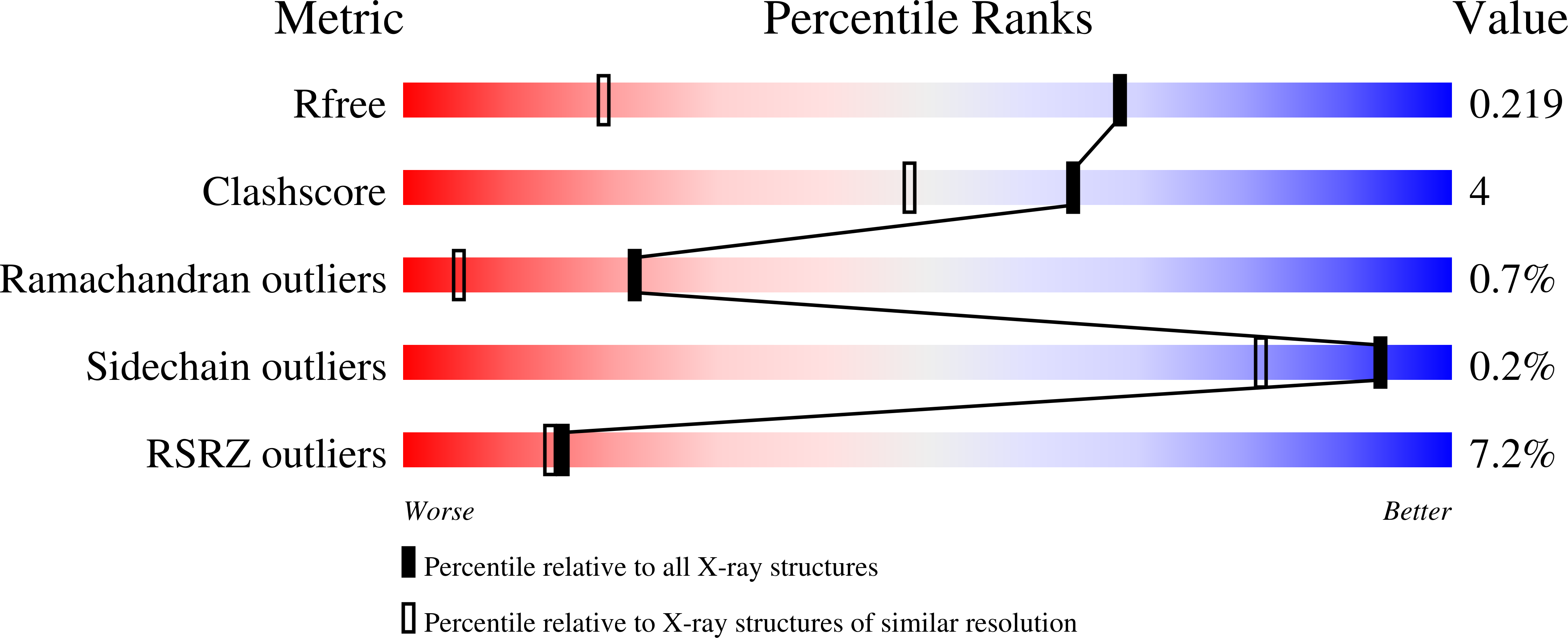
Resolution:
1.40 Å
R-Value Free:
0.21
R-Value Work:
0.17
R-Value Observed:
0.17
Space Group:
P 1 21 1
Macromolecular Entities
Polymer Type:polypeptide(L)
Molecule:Sensor protein
Gene (Uniprot):cusS
Chain IDs:A, B, C, D, E, F
Chain Length:217
Number of Molecules:6
Biological Source:Escherichia coli 'BL21-Gold(DE3)pLysS AG'
Ligand Molecules
Primary Citation
Crystal structure of the Escherichia coli CusS kinase core.
J.Struct.Biol. 214 107883 107883 (2022)
PMID: 35907487 DOI: 10.1016/j.jsb.2022.107883

Abstact

The CusS histidine kinase is a member of Escherichia coli two-component signal transduction system, engaged in a response to copper ions excess in the cell periplasm. The periplasmic sensor domain of CusS binds the free copper ions and the CusS kinase core phosphorylates the cognate CusR which regulates transcription of the efflux pomp CusCBA. A small amount of copper ions is indispensable for the aerobic cell metabolism. Nonetheless, its excess in the cytoplasm generates damaging and reactive hydroxyl radicals. For that reason, understanding the bacterial copper sensing mechanisms can contribute to reducing bacterial copper-resistance and developing bactericidal copper-based materials. The crystal structure of the CusS kinase core was solved at the resolution of 1.4 Å. The cytoplasmic catalytic core domains formed a homodimer. Based on the obtained structure, the intramolecular and intermolecular interactions crucial for the mechanism of CusS autophosphorylation were described.

Legend

Protein

Chemical

Disease

Primary Citation of related structures