Deposition Date
2022-04-03
Release Date
2022-10-12
Last Version Date
2024-11-13
Entry Detail
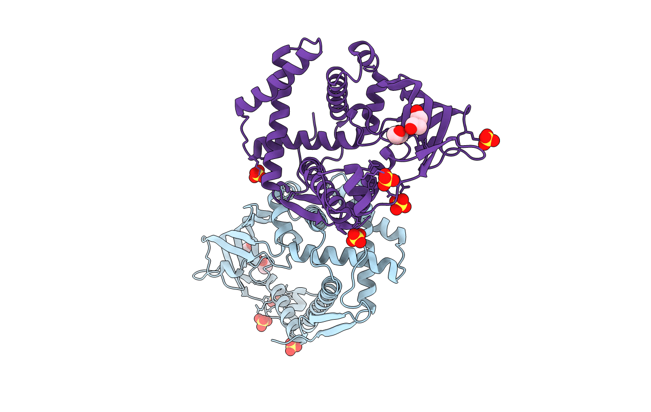
Biological Source:
Source Organism(s):
Chlamydomonas reinhardtii (Taxon ID: 3055)
Expression System(s):
Method Details:
Experimental Method:
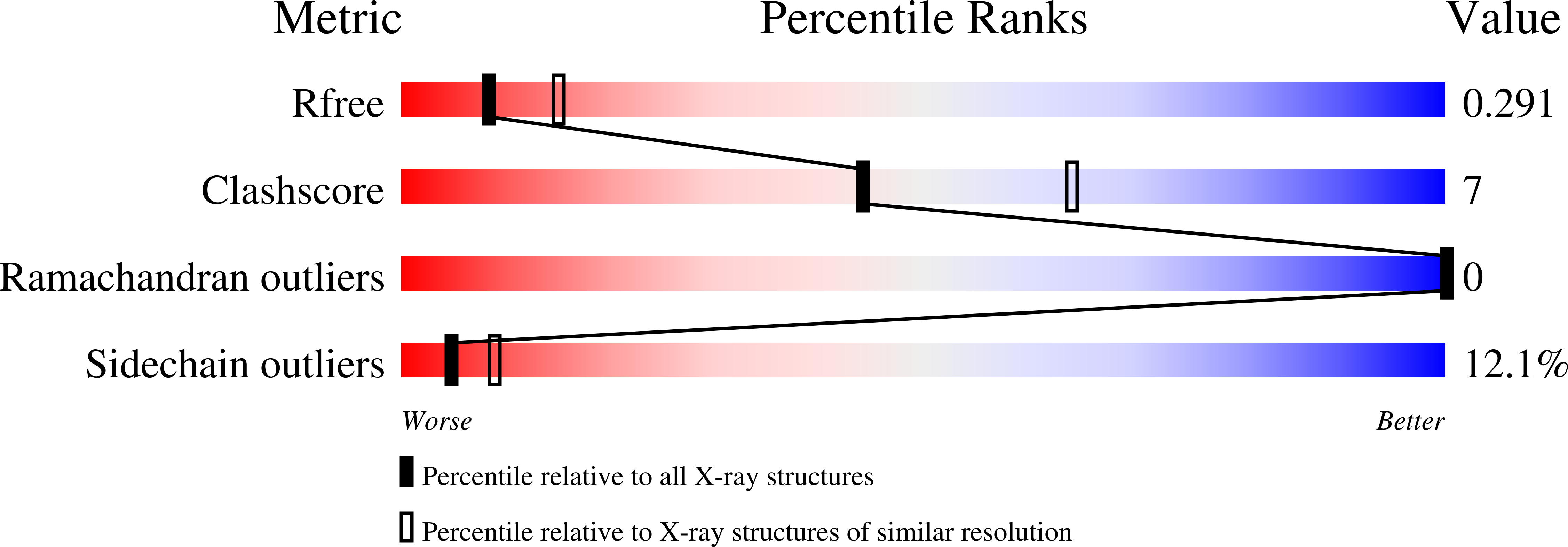
Resolution:
2.60 Å
R-Value Free:
0.28
R-Value Work:
0.23
R-Value Observed:
0.23
Space Group:
C 1 2 1