
Deposition Date
2022-03-24
Release Date
2022-06-29
Last Version Date
2024-06-19
Entry Detail
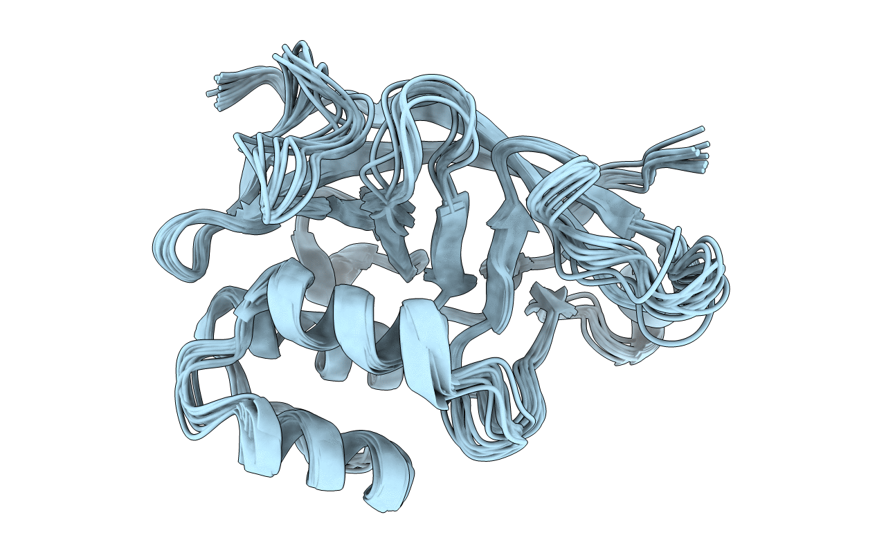
PDB ID:
7ZBQ
Keywords:
Title:
Structure of the ADP-ribosyltransferase TccC3HVR from Photorhabdus Luminescens
Biological Source:
Source Organism(s):
Photorhabdus luminescens (Taxon ID: 29488)
Expression System(s):
Method Details:
Experimental Method:
Conformers Calculated:
400
Conformers Submitted:
10
Selection Criteria:
structures with the lowest energy
