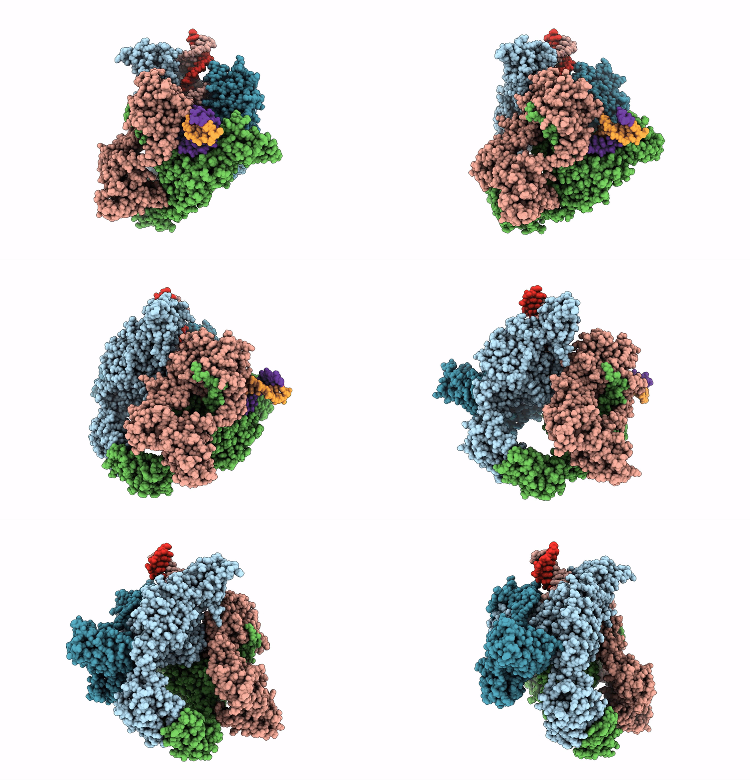
7Z9K image
Entry Detail
PDB ID:
7Z9K
EMDB ID:
Keywords:
Title:
E.coli gyrase holocomplex with 217 bp DNA and Albi-1 (site TG)
Biological Source:
PDB Version:
Deposition Date:
2022-03-21
Release Date:
2023-03-08
Method Details:
Experimental Method:
Resolution:
3.25 Å
Aggregation State:
PARTICLE
Reconstruction Method:
SINGLE PARTICLE
Macromolecular Entities
Polymer Type:polypeptide(L)
Description:DNA gyrase subunit A
Chain IDs:A, C
Chain Length:881
Number of Molecules:2
Biological Source:Escherichia coli str. K-12 substr. MG1655
Polymer Type:polypeptide(L)
Description:DNA gyrase subunit B
Chain IDs:B, D
Chain Length:811
Number of Molecules:2
Biological Source:Escherichia coli str. K-12 substr. MG1655
Polymer Type:polydeoxyribonucleotide
Description:DNA (5'-D(P*AP*AP*AP*TP*CP*TP*GP*TP*GP*CP*GP*GP*GP*T)-3')
Chain IDs:E
Chain Length:14
Number of Molecules:1
Biological Source:Escherichia phage Mu
Polymer Type:polydeoxyribonucleotide
Description:DNA (5'-D(P*AP*GP*AP*AP*TP*CP*AP*GP*GP*CP*AP*TP*AP*A)-3')
Chain IDs:F
Chain Length:14
Number of Molecules:1
Biological Source:Escherichia phage Mu
Polymer Type:polydeoxyribonucleotide
Description:DNA (5'-D(*AP*AP*TP*CP*AP*CP*CP*CP*GP*CP*AP*CP*AP*GP*AP*TP*TP*T)-3')
Chain IDs:G
Chain Length:18
Number of Molecules:1
Biological Source:Escherichia phage Mu
Polymer Type:polydeoxyribonucleotide
Description:DNA (5'-D(*GP*AP*TP*TP*TP*TP*AP*TP*GP*CP*CP*TP*GP*AP*TP*TP*CP*T)-3')
Chain IDs:H
Chain Length:18
Number of Molecules:1
Biological Source:Escherichia phage Mu
Modified Residue
Compound ID Chain ID Parent Comp ID Details 2D Image
PTR A TYR modified residue
Primary Citation
Molecular mechanism of topoisomerase poisoning by the peptide antibiotic albicidin.
Nat Catal 6 52 67 (2023)
PMID: 36741192 DOI: 10.1038/s41929-022-00904-1

Abstact

The peptide antibiotic albicidin is a DNA topoisomerase inhibitor with low-nanomolar bactericidal activity towards fluoroquinolone-resistant Gram-negative pathogens. However, its mode of action is poorly understood. We determined a 2.6 Å resolution cryoelectron microscopy structure of a ternary complex between Escherichia coli topoisomerase DNA gyrase, a 217 bp double-stranded DNA fragment and albicidin. Albicidin employs a dual binding mechanism where one end of the molecule obstructs the crucial gyrase dimer interface, while the other intercalates between the fragments of cleaved DNA substrate. Thus, albicidin efficiently locks DNA gyrase, preventing it from religating DNA and completing its catalytic cycle. Two additional structures of this trapped state were determined using synthetic albicidin analogues that demonstrate improved solubility, and activity against a range of gyrase variants and E. coli topoisomerase IV. The extraordinary promiscuity of the DNA-intercalating region of albicidins and their excellent performance against fluoroquinolone-resistant bacteria holds great promise for the development of last-resort antibiotics.

Legend

Protein

Chemical

Disease

Primary Citation of related structures