7Z6E image
Entry Detail
PDB ID:
7Z6E
Title:
Structure of the C1-PH-CNH regulatory module of MRCK1
Biological Source:
Source Organism:
Host Organism:
PDB Version:
Deposition Date:
2022-03-11
Release Date:
2023-02-22
Method Details:
Experimental Method:
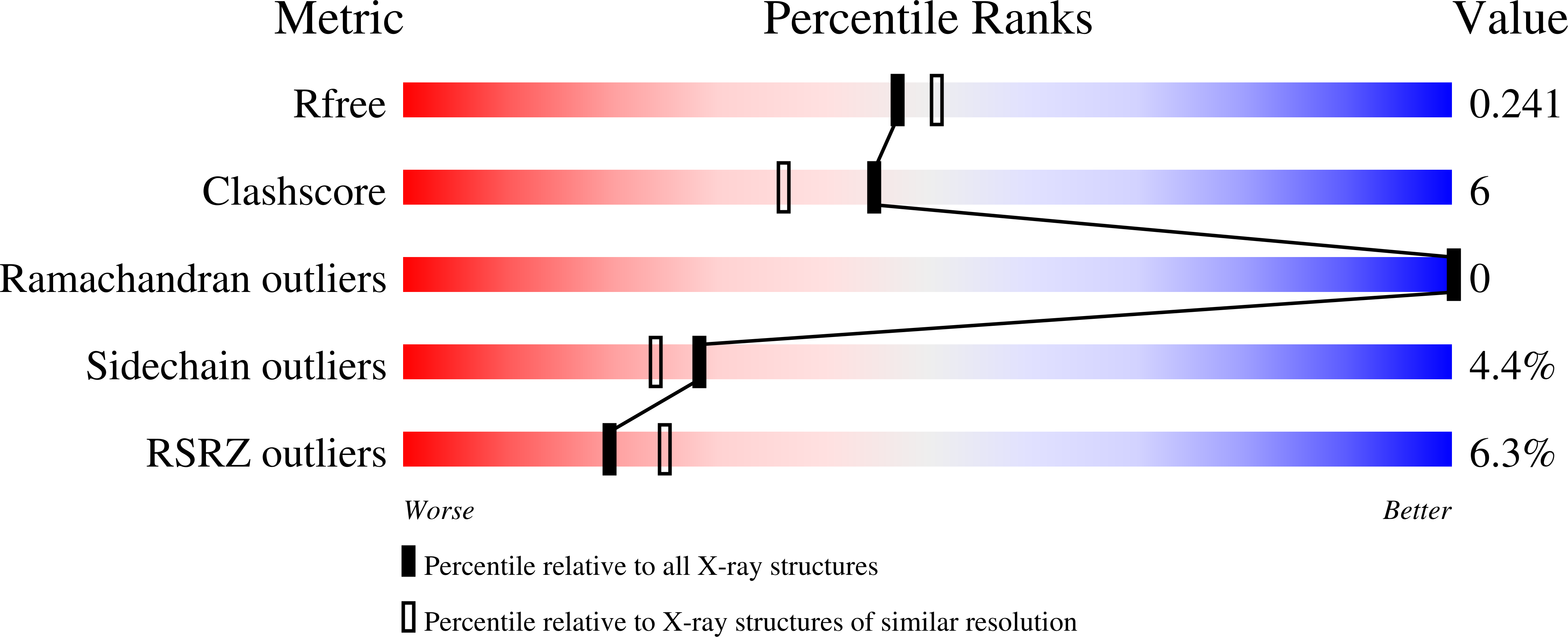
Resolution:
2.14 Å
R-Value Free:
0.24
R-Value Work:
0.19
R-Value Observed:
0.19
Space Group:
P 1
Macromolecular Entities
Polymer Type:polypeptide(L)
Description:Serine/threonine-protein kinase mrck-1
Chain IDs:A, B, C, D, E
Chain Length:582
Number of Molecules:5
Biological Source:Caenorhabditis elegans
Ligand Molecules
Primary Citation
Structure and regulation of the myotonic dystrophy kinase-related Cdc42-binding kinase.
Structure 31 435 ? (2023)
PMID: 36854301 DOI: 10.1016/j.str.2023.02.002

Abstact

Protein kinases of the dystonia myotonica protein kinase (DMPK) family are critical regulators of actomyosin contractility in cells. The DMPK kinase MRCK1 is required for the activation of myosin, leading to the development of cortical tension, apical constriction, and early gastrulation. Here, we present the structure, conformation, and membrane-binding properties of Caenorhabditis elegans MRCK1. MRCK1 forms a homodimer with N-terminal kinase domains, a parallel coiled coil of 55 nm, and a C-terminal tripartite module of C1, pleckstrin homology (PH), and citron homology (CNH) domains. We report the high-resolution structure of the membrane-binding C1-PH-CNH module of MRCK1 and, using high-throughput and conventional liposome-binding assays, determine its binding to specific phospholipids. We further characterize the interaction of the C-terminal CRIB motif with Cdc42. The length of the coiled-coil domain of DMPK kinases is remarkably conserved over millions of years of evolution, suggesting that they may function as molecular rulers to position kinase activity at a fixed distance from the membrane.

Legend

Protein

Chemical

Disease

Primary Citation of related structures