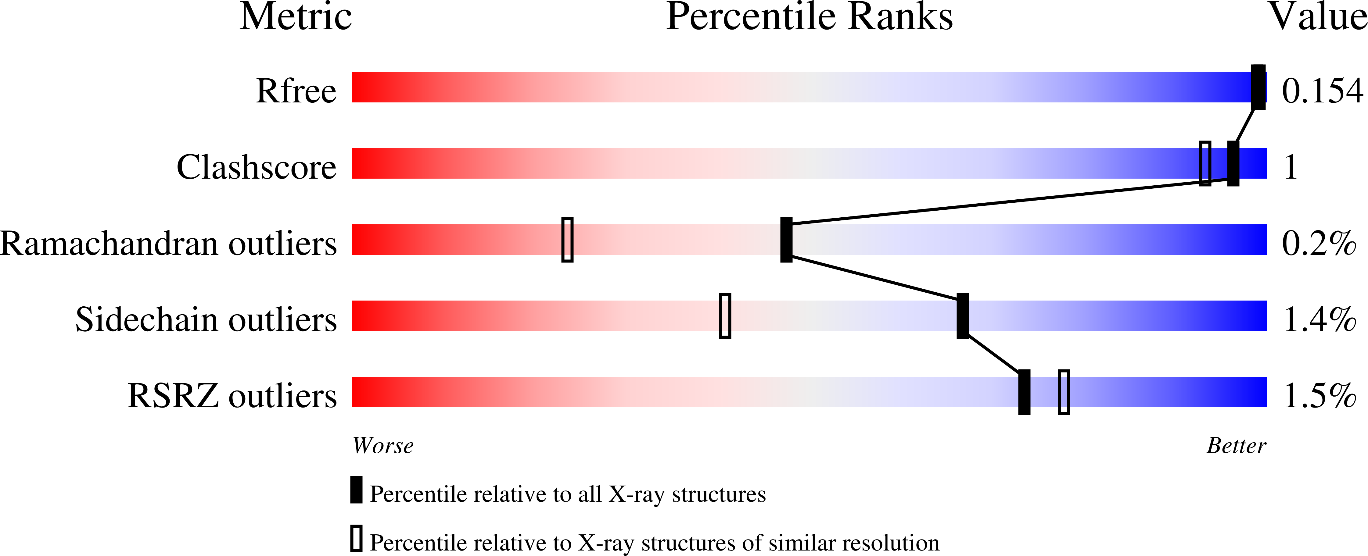
7Z5O image
Deposition Date 2022-03-09
Release Date 2022-07-13
Last Version Date 2024-11-13
Entry Detail
PDB ID:
7Z5O
Keywords:
Title:
W-formate dehydrogenase from Desulfovibrio vulgaris - Dithionite reduced form
Biological Source:
Method Details:
Experimental Method:
Resolution:
1.53 Å
R-Value Free:
0.15
R-Value Work:
0.11
Space Group:
P 21 21 21
Macromolecular Entities
Structures with similar UniProt ID
Protein Blast
Polymer Type:polypeptide(L)
Molecule:Formate dehydrogenase, alpha subunit, selenocysteine-containing
Gene (Uniprot):fdnG-1
Chain IDs:A (auth: AAA)
Chain Length:1009
Number of Molecules:1
Biological Source:Desulfovibrio vulgaris str. Hildenborough
Structures with similar UniProt ID
Protein Blast
Polymer Type:polypeptide(L)
Molecule:Formate dehydrogenase, beta subunit, putative
Gene (Uniprot):DVU_0588
Chain IDs:B (auth: BBB)
Chain Length:236
Number of Molecules:1
Biological Source:Desulfovibrio vulgaris str. Hildenborough
Primary Citation
Spectroscopic and Structural Characterization of Reduced Desulfovibrio vulgaris Hildenborough W-FdhAB Reveals Stable Metal Coordination during Catalysis.
Acs Chem.Biol. 17 1901 1909 (2022)
PMID: 35766974 DOI: 10.1021/acschembio.2c00336

Abstact

Metal-dependent formate dehydrogenases are important enzymes due to their activity of CO2 reduction to formate. The tungsten-containing FdhAB formate dehydrogenase from Desulfovibrio vulgaris Hildenborough is a good example displaying high activity, simple composition, and a notable structural and catalytic robustness. Here, we report the first spectroscopic redox characterization of FdhAB metal centers by EPR. Titration with dithionite or formate leads to reduction of three [4Fe-4S]1+ clusters, and full reduction requires Ti(III)-citrate. The redox potentials of the four [4Fe-4S]1+ centers range between -250 and -530 mV. Two distinct WV signals were detected, WDV and WFV, which differ in only the g2-value. This difference can be explained by small variations in the twist angle of the two pyranopterins, as determined through DFT calculations of model compounds. The redox potential of WVI/V was determined to be -370 mV when reduced by dithionite and -340 mV when reduced by formate. The crystal structure of dithionite-reduced FdhAB was determined at high resolution (1.5 Å), revealing the same structural alterations as reported for the formate-reduced structure. These results corroborate a stable six-ligand W coordination in the catalytic intermediate WV state of FdhAB.

Legend

Protein

Chemical

Disease

Primary Citation of related structures