Deposition Date
2022-03-02
Release Date
2023-09-13
Last Version Date
2024-02-28
Entry Detail
PDB ID:
7Z3F
Keywords:
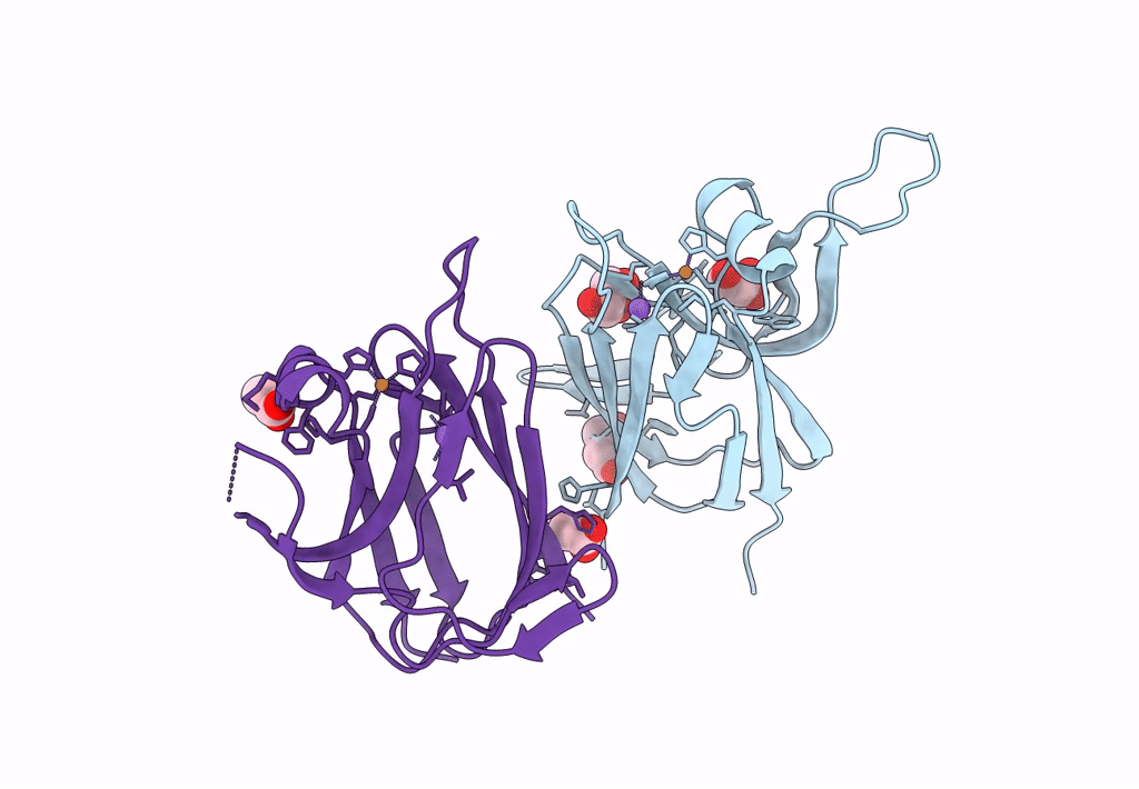
Title:
Crystal structure of the cupredoxin AcoP from Acidithiobacillus ferrooxidans, oxidized form
Biological Source:
Source Organism(s):
Acidithiobacillus ferrooxidans (Taxon ID: 920)
Expression System(s):
Method Details:
Experimental Method:
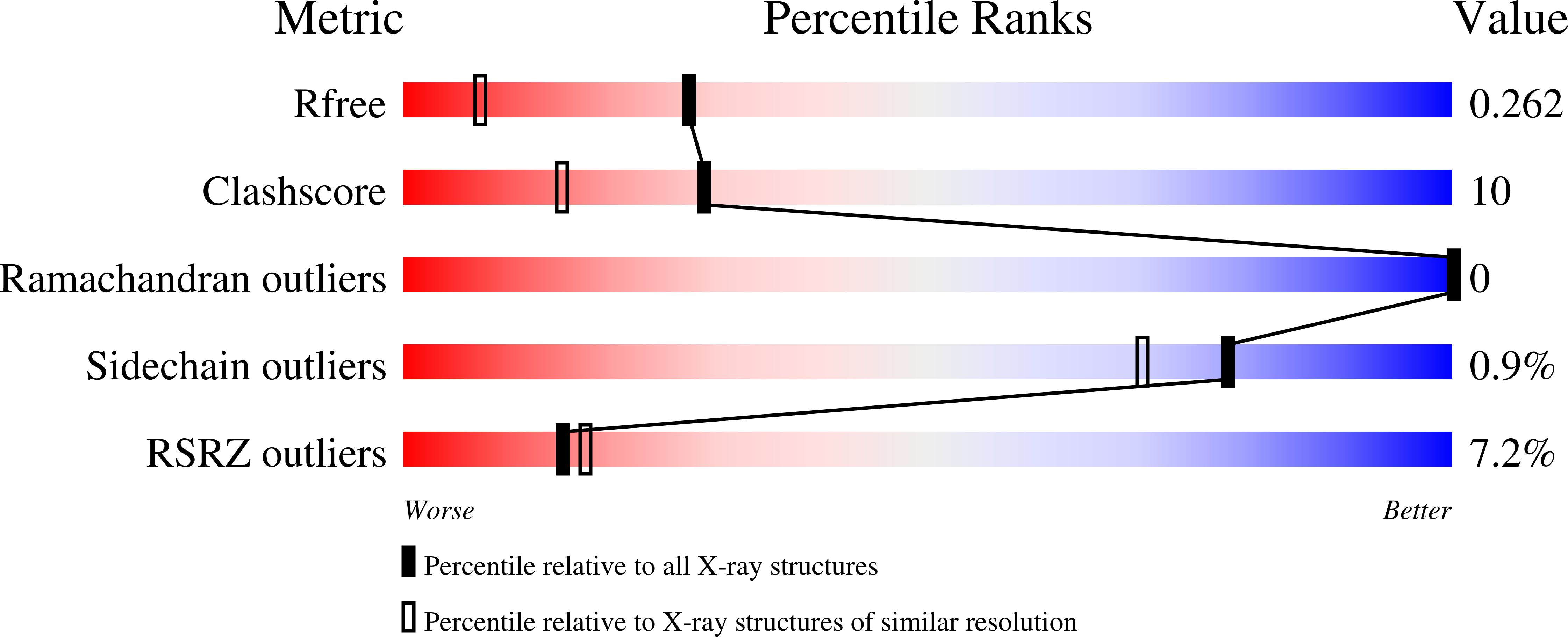
Resolution:
1.70 Å
R-Value Free:
0.26
R-Value Work:
0.23
R-Value Observed:
0.23
Space Group:
P 41 21 2