7YWP image
Deposition Date 2022-02-14
Release Date 2023-02-22
Last Version Date 2024-11-13
Entry Detail
PDB ID:
7YWP
Keywords:
Title:
Closed conformation of Oligopeptidase B from Serratia proteomaculans with covalently bound TCK
Biological Source:
Source Organism:
Host Organism:
Method Details:
Experimental Method:
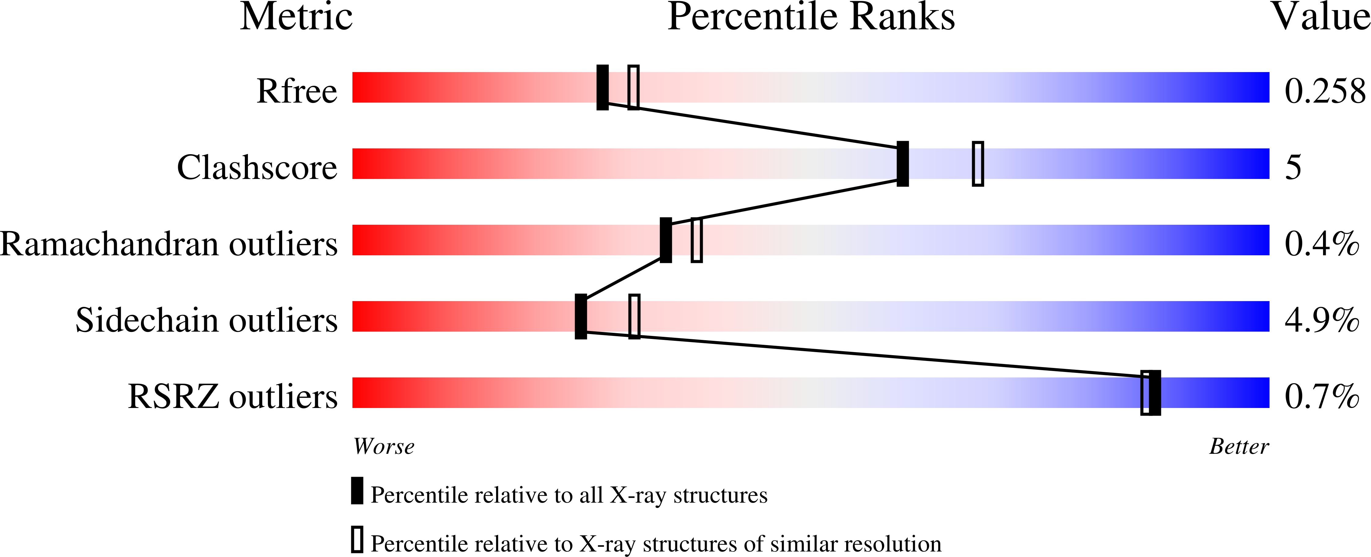
Resolution:
2.20 Å
R-Value Free:
0.25
R-Value Work:
0.18
R-Value Observed:
0.18
Space Group:
P 21 21 21
Macromolecular Entities
Structures with similar UniProt ID
Protein Blast
Polymer Type:polypeptide(L)
Molecule:Oligopeptidase B
Gene (Uniprot):opdB
Chain IDs:A
Chain Length:676
Number of Molecules:1
Biological Source:Serratia proteamaculans
Ligand Molecules
Primary Citation
Crystal Structure of Inhibitor-Bound Bacterial Oligopeptidase B in the Closed State: Similarity and Difference between Protozoan and Bacterial Enzymes.
Int J Mol Sci 24 ? ? (2023)
PMID: 36768612 DOI: 10.3390/ijms24032286

Abstact

The crystal structure of bacterial oligopeptidase B from Serratia proteamaculans (SpOpB) in complex with a chloromethyl ketone inhibitor was determined at 2.2 Å resolution. SpOpB was crystallized in a closed (catalytically active) conformation. A single inhibitor molecule bound simultaneously to the catalytic residues S532 and H652 mimicked a tetrahedral intermediate of the catalytic reaction. A comparative analysis of the obtained structure and the structure of OpB from Trypanosoma brucei (TbOpB) in a closed conformation showed that in both enzymes, the stabilization of the D-loop (carrying the catalytic D) in a position favorable for the formation of a tetrahedral complex occurs due to interaction with the neighboring loop from the β-propeller. However, the modes of interdomain interactions were significantly different for bacterial and protozoan OpBs. Instead of a salt bridge (as in TbOpB), in SpOpB, a pair of polar residues following the catalytic D617 and a pair of neighboring arginine residues from the β-propeller domain formed complementary oppositely charged surfaces. Bioinformatics analysis and structural modeling show that all bacterial OpBs can be divided into two large groups according to these two modes of D-loop stabilization in closed conformations.

Legend

Protein

Chemical

Disease

Primary Citation of related structures
Feedback Form
Name
Email
Institute
Feedback