7YUJ image
Deposition Date 2022-08-17
Release Date 2023-08-30
Last Version Date 2024-03-13
Entry Detail
PDB ID:
7YUJ
Keywords:
Title:
Crystal structure of HOIL-1L(365-510)
Biological Source:
Source Organism:
Homo sapiens (Taxon ID: 9606)
Host Organism:
Method Details:
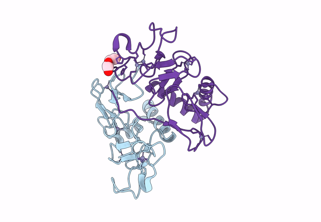
Experimental Method:
Resolution:
1.87 Å
R-Value Free:
0.18
R-Value Work:
0.15
R-Value Observed:
0.15
Space Group:
P 1 21 1
Macromolecular Entities
Protein Blast
Polymer Type:polypeptide(L)
Molecule:RanBP-type and C3HC4-type zinc finger-containing protein 1
Gene (Uniprot):RBCK1
Chain IDs:A, B
Chain Length:150
Number of Molecules:2
Biological Source:Homo sapiens
Primary Citation
Mechanistic insights into the enzymatic activity of E3 ligase HOIL-1L and its regulation by the linear ubiquitin chain binding.
Sci Adv 9 eadi4599 eadi4599 (2023)
PMID: 37831767 DOI: 10.1126/sciadv.adi4599

Abstact

Heme-oxidized IRP2 ubiquitin ligase 1 (HOIL-1L) serves as a unique E3 ligase to catalyze the mono-ubiquitination of relevant protein or sugar substrates and plays vital roles in numerous cellular processes in mammals. However, the molecular mechanism underpinning the E3 activity of HOIL-1L and the related regulatory mechanism remain elusive. Here, we report the crystal structure of the catalytic core region of HOIL-1L and unveil the key catalytic triad residues of HOIL-1L. Moreover, we discover that HOIL-1L contains two distinct linear di-ubiquitin binding sites that can synergistically bind to linear tetra-ubiquitin, and the binding of HOIL-1L with linear tetra-ubiquitin can promote its E3 activity. The determined HOIL-1L/linear tetra-ubiquitin complex structure not only elucidates the detailed binding mechanism of HOIL-1L with linear tetra-ubiquitin but also uncovers a unique allosteric ubiquitin-binding site for the activation of HOIL-1L. In all, our findings provide mechanistic insights into the E3 activity of HOIL-1L and its regulation by the linear ubiquitin chain binding.

Legend

Protein

Chemical

Disease

Primary Citation of related structures