
Deposition Date
2022-08-08
Release Date
2023-03-29
Last Version Date
2025-07-02
Entry Detail
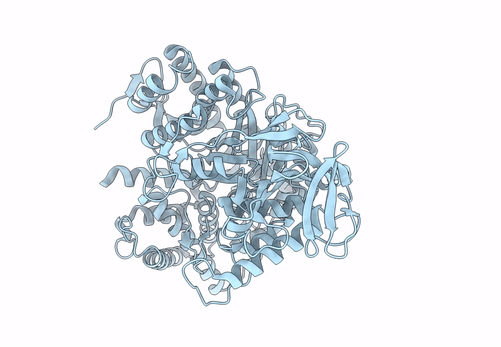
PDB ID:
7YQM
Keywords:
Title:
2.9-angstrom cryo-EM structure of Ecoli malate synthase G
Biological Source:
Source Organism(s):
Escherichia coli DH5[alpha] (Taxon ID: 668369)
Expression System(s):
Method Details:
Experimental Method:
Resolution:
2.89 Å
Aggregation State:
PARTICLE
Reconstruction Method:
SINGLE PARTICLE
