7YQ2 image
Entry Detail
PDB ID:
7YQ2
Title:
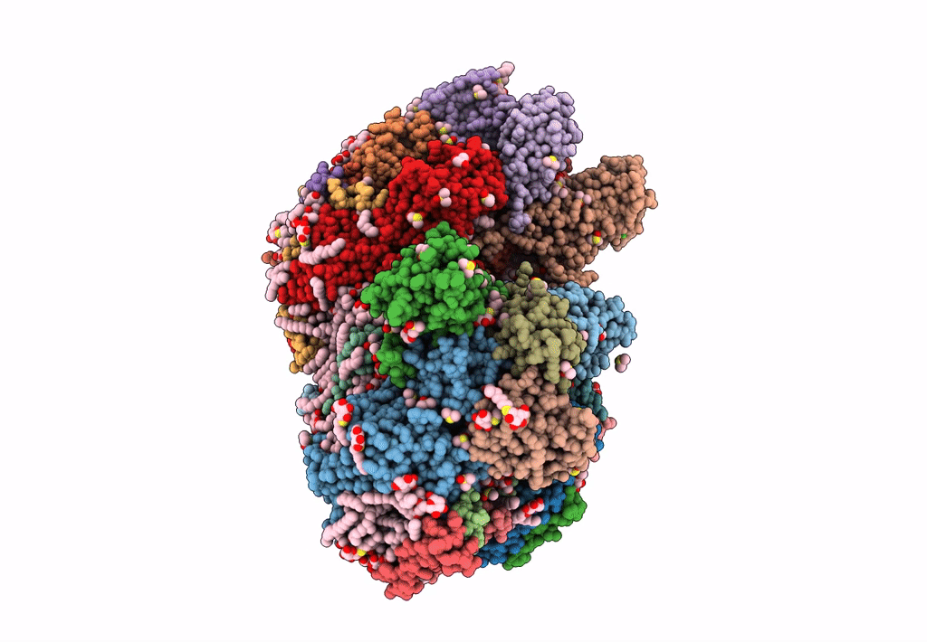
Crystal structure of photosystem II expressing psbA2 gene only
Biological Source:
PDB Version:
Deposition Date:
2022-08-05
Release Date:
2022-11-30
Method Details:
Experimental Method:
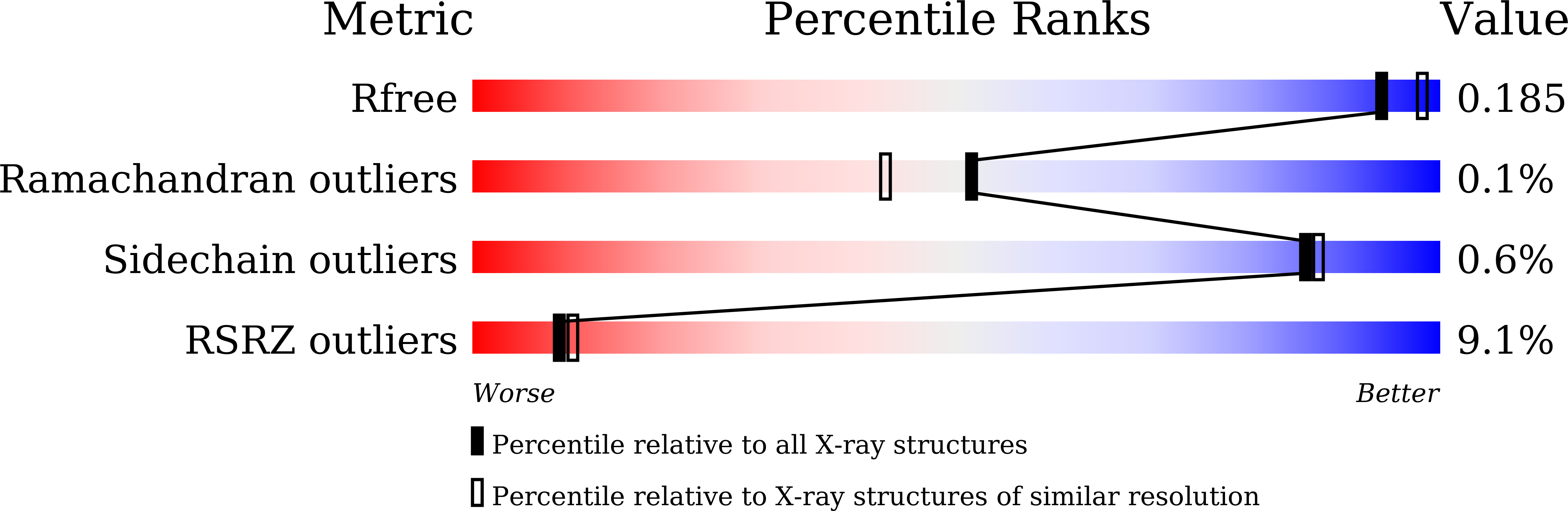
Resolution:
1.90 Å
R-Value Free:
0.18
R-Value Work:
0.15
R-Value Observed:
0.15
Space Group:
P 21 21 21
Macromolecular Entities
Polymer Type:polypeptide(L)
Description:Photosystem II protein D1 2
Chain IDs:A, T (auth: a)
Chain Length:360
Number of Molecules:2
Biological Source:Thermosynechococcus vestitus BP-1
Polymer Type:polypeptide(L)
Description:Photosystem II CP47 reaction center protein
Chain IDs:B, U (auth: b)
Chain Length:510
Number of Molecules:2
Biological Source:Thermosynechococcus vestitus BP-1
Polymer Type:polypeptide(L)
Description:Photosystem II CP43 reaction center protein
Chain IDs:C, V (auth: c)
Chain Length:461
Number of Molecules:2
Biological Source:Thermosynechococcus vestitus BP-1
Polymer Type:polypeptide(L)
Description:Photosystem II D2 protein
Chain IDs:D, W (auth: d)
Chain Length:352
Number of Molecules:2
Biological Source:Thermosynechococcus vestitus BP-1
Polymer Type:polypeptide(L)
Description:Cytochrome b559 subunit alpha
Chain IDs:E, X (auth: e)
Chain Length:84
Number of Molecules:2
Biological Source:Thermosynechococcus vestitus BP-1
Polymer Type:polypeptide(L)
Description:Cytochrome b559 subunit beta
Chain IDs:F, Y (auth: f)
Chain Length:45
Number of Molecules:2
Biological Source:Thermosynechococcus vestitus BP-1
Polymer Type:polypeptide(L)
Description:Photosystem II reaction center protein H
Chain IDs:G (auth: H), Z (auth: h)
Chain Length:66
Number of Molecules:2
Biological Source:Thermosynechococcus vestitus BP-1
Polymer Type:polypeptide(L)
Description:Photosystem II reaction center protein I
Chain IDs:H (auth: I), AA (auth: i)
Chain Length:38
Number of Molecules:2
Biological Source:Thermosynechococcus vestitus BP-1
Polymer Type:polypeptide(L)
Description:Photosystem II reaction center protein J
Chain IDs:I (auth: J), BA (auth: j)
Chain Length:40
Number of Molecules:2
Biological Source:Thermosynechococcus vestitus BP-1
Polymer Type:polypeptide(L)
Description:Photosystem II reaction center protein K
Chain IDs:J (auth: K), CA (auth: k)
Chain Length:46
Number of Molecules:2
Biological Source:Thermosynechococcus vestitus BP-1
Polymer Type:polypeptide(L)
Description:Photosystem II reaction center protein L
Chain IDs:K (auth: L), DA (auth: l)
Chain Length:37
Number of Molecules:2
Biological Source:Thermosynechococcus vestitus BP-1
Polymer Type:polypeptide(L)
Description:Photosystem II reaction center protein M
Chain IDs:L (auth: M), EA (auth: m)
Chain Length:36
Number of Molecules:2
Biological Source:Thermosynechococcus vestitus BP-1
Polymer Type:polypeptide(L)
Description:Photosystem II manganese-stabilizing polypeptide
Chain IDs:M (auth: O), FA (auth: o)
Chain Length:272
Number of Molecules:2
Biological Source:Thermosynechococcus vestitus BP-1
Polymer Type:polypeptide(L)
Description:Photosystem II protein Y
Chain IDs:MA (auth: R)
Chain Length:41
Number of Molecules:1
Biological Source:Thermosynechococcus vestitus BP-1
Polymer Type:polypeptide(L)
Description:Photosystem II reaction center protein T
Chain IDs:N (auth: T), GA (auth: t)
Chain Length:32
Number of Molecules:2
Biological Source:Thermosynechococcus vestitus BP-1
Polymer Type:polypeptide(L)
Description:Photosystem II 12 kDa extrinsic protein
Chain IDs:O (auth: U), HA (auth: u)
Chain Length:134
Number of Molecules:2
Biological Source:Thermosynechococcus vestitus BP-1
Polymer Type:polypeptide(L)
Description:Cytochrome c-550
Chain IDs:P (auth: V), IA (auth: v)
Chain Length:163
Number of Molecules:2
Biological Source:Thermosynechococcus vestitus BP-1
Polymer Type:polypeptide(L)
Description:Photosystem II reaction center X protein
Chain IDs:R (auth: X), KA (auth: x)
Chain Length:41
Number of Molecules:2
Biological Source:Thermosynechococcus vestitus BP-1
Polymer Type:polypeptide(L)
Description:Photosystem II reaction center protein Ycf12
Chain IDs:Q (auth: Y), JA (auth: y)
Chain Length:46
Number of Molecules:2
Biological Source:Thermosynechococcus vestitus BP-1
Polymer Type:polypeptide(L)
Description:Photosystem II reaction center protein Z
Chain IDs:S (auth: Z), LA (auth: z)
Chain Length:62
Number of Molecules:2
Biological Source:Thermosynechococcus vestitus BP-1
Modified Residue
Compound ID Chain ID Parent Comp ID Details 2D Image
FME H MET modified residue
Primary Citation
Crystal structures of photosystem II from a cyanobacterium expressing psbA 2 in comparison to psbA 3 reveal differences in the D1 subunit.
J.Biol.Chem. 298 102668 102668 (2022)
PMID: 36334624 DOI: 10.1016/j.jbc.2022.102668

Abstact

Three psbA genes (psbA1, psbA2, and psbA3) encoding the D1 subunit of photosystem II (PSII) are present in the thermophilic cyanobacterium Thermosynechococcus elongatus and are expressed differently in response to changes in the growth environment. To clarify the functional differences of the D1 protein expressed from these psbA genes, PSII dimers from two strains, each expressing only one psbA gene (psbA2 or psbA3), were crystallized, and we analyzed their structures at resolutions comparable to previously studied PsbA1-PSII. Our results showed that the hydrogen bond between pheophytin/D1 (PheoD1) and D1-130 became stronger in PsbA2- and PsbA3-PSII due to change of Gln to Glu, which partially explains the increase in the redox potential of PheoD1 observed in PsbA3. In PsbA2, one hydrogen bond was lost in PheoD1 due to the change of D1-Y147F, which may explain the decrease in stability of PheoD1 in PsbA2. Two water molecules in the Cl-1 channel were lost in PsbA2 due to the change of D1-P173M, leading to the narrowing of the channel, which may explain the lower efficiency of the S-state transition beyond S2 in PsbA2-PSII. In PsbA3-PSII, a hydrogen bond between D1-Ser270 and a sulfoquinovosyl-diacylglycerol molecule near QB disappeared due to the change of D1-Ser270 in PsbA1 and PsbA2 to D1-Ala270. This may result in an easier exchange of bound QB with free plastoquinone, hence an enhancement of oxygen evolution in PsbA3-PSII due to its high QB exchange efficiency. These results provide a structural basis for further functional examination of the three PsbA variants.

Legend

Protein

Chemical

Disease

Primary Citation of related structures