7YOB image
Deposition Date 2022-08-01
Release Date 2022-08-17
Last Version Date 2023-11-29
Entry Detail
PDB ID:
7YOB
Keywords:
Title:
Crystal structure of Aldehyde dehydrogenase 1A1 from mouse
Biological Source:
Source Organism:
Mus musculus (Taxon ID: 10090)
Host Organism:
Method Details:
Experimental Method:
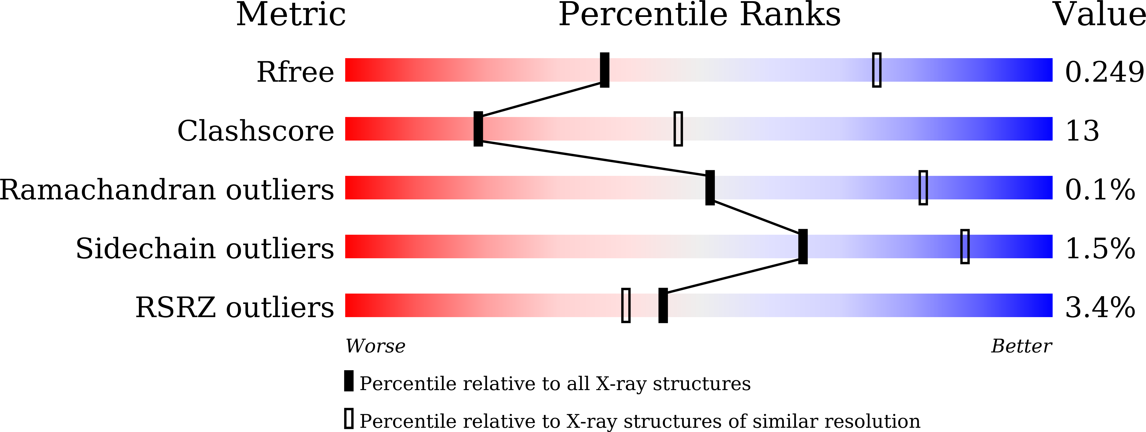
Resolution:
2.89 Å
R-Value Free:
0.24
R-Value Work:
0.19
R-Value Observed:
0.19
Space Group:
P 1 21 1
Macromolecular Entities
Structures with similar UniProt ID
Protein Blast
Polymer Type:polypeptide(L)
Molecule:Aldehyde dehydrogenase 1A1
Gene (Uniprot):Aldh1a1
Chain IDs:A, B, C, D, E, F (auth: G), G (auth: H), H (auth: F)
Chain Length:507
Number of Molecules:8
Biological Source:Mus musculus
Ligand Molecules
Primary Citation
Crystal structure of aldehyde dehydrogenase 1A1 from mouse.
Biochem.Biophys.Res.Commun. 628 141 146 (2022)
PMID: 36084552 DOI: 10.1016/j.bbrc.2022.08.054

Abstact

Aldehyde dehydrogenase 1A1 (ALDH1A1) is an enzyme that catalyzes the NAD+-dependent oxidation of aldehydes to carboxylic acids, participating in various metabolic processes. Currently, only structures from human and Ovis aries have been reported. Here we show a 2.89 Å resolution structure of ALDH1A1 from mice using X-ray crystallography. We performed a detailed analysis of the structure and compared it with ALDH1A1 structures from two other species, highlighting the significance of the differences. Structural superimposition reveals that the tetrameric molecule is asymmetrical, and the NAD+-binding domain exhibits a certain rotation. In addition, the noticeable structural differences were detected, including the unique contact between Ser461 and Asp148, as well as the side chain orientations of three amino acids residues, Asn474, Met471 and Phe466. This study helps to expand the structural diversity of the ALDH family.

Legend

Protein

Chemical

Disease

Primary Citation of related structures