7YNU image
Deposition Date 2022-08-01
Release Date 2022-09-21
Last Version Date 2024-11-06
Entry Detail
PDB ID:
7YNU
Title:
Crystal structure of Hen Egg white LYSOZYME introduced with O-(2-nitrobenzyl)-L-tyrosine
Biological Source:
Source Organism:
Gallus gallus (Taxon ID: 9031)
Host Organism:
Method Details:
Experimental Method:
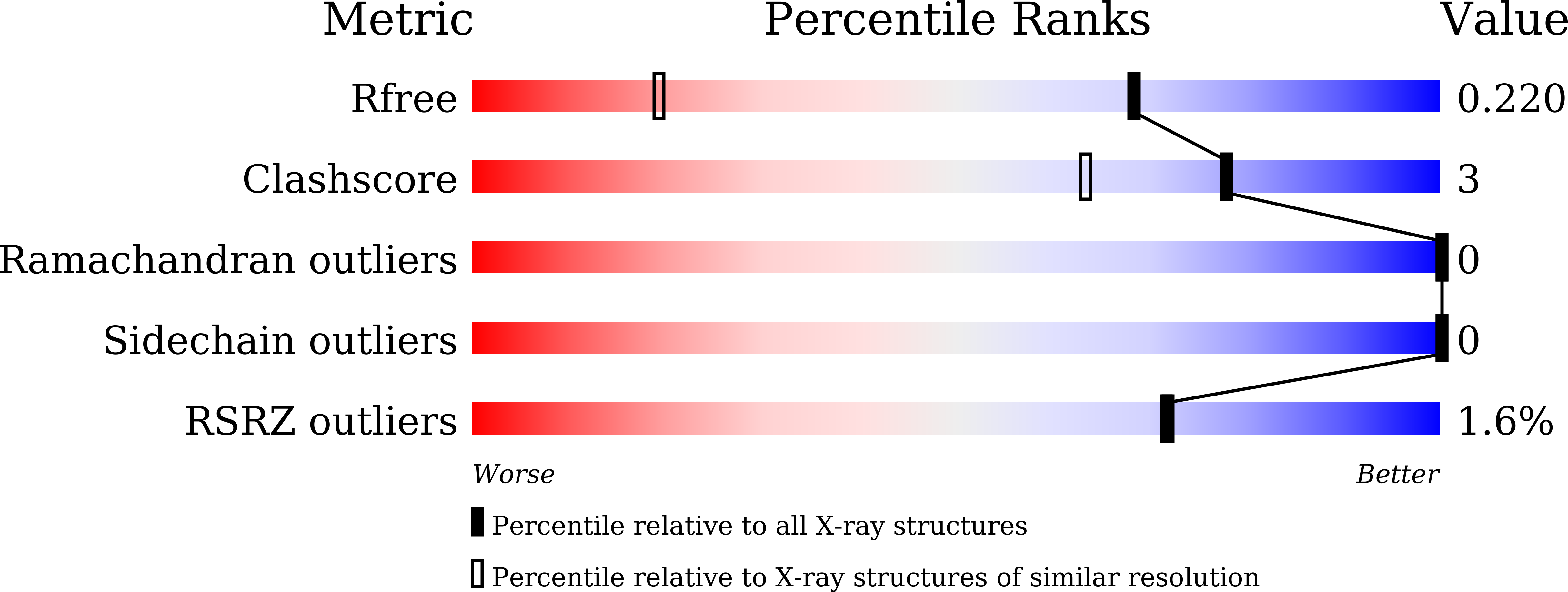
Resolution:
1.44 Å
R-Value Free:
0.22
R-Value Work:
0.19
R-Value Observed:
0.19
Space Group:
P 43 21 2
Macromolecular Entities
Structures with similar UniProt ID
Protein Blast
Polymer Type:polypeptide(L)
Molecule:Lysozyme C
Gene (Uniprot):LYZ
Chain IDs:A
Chain Length:129
Number of Molecules:1
Biological Source:Gallus gallus
Modified Residue
Compound ID Chain ID Parent Comp ID Details 2D Image
J2F A TYR modified residue
Primary Citation
Crystal Structure of an Archaeal Tyrosyl-tRNA Synthetase Bound to Photocaged L-Tyrosine and Its Potential Application to Time-Resolved X-ray Crystallography.
Int J Mol Sci 23 ? ? (2022)
PMID: 36142308 DOI: 10.3390/ijms231810399

Abstact

Genetically encoded caged amino acids can be used to control the dynamics of protein activities and cellular localization in response to external cues. In the present study, we revealed the structural basis for the recognition of O-(2-nitrobenzyl)-L-tyrosine (oNBTyr) by its specific variant of Methanocaldococcus jannaschii tyrosyl-tRNA synthetase (oNBTyrRS), and then demonstrated its potential availability for time-resolved X-ray crystallography. The substrate-bound crystal structure of oNBTyrRS at a 2.79 Å resolution indicated that the replacement of tyrosine and leucine at positions 32 and 65 by glycine (Tyr32Gly and Leu65Gly, respectively) and Asp158Ser created sufficient space for entry of the bulky substitute into the amino acid binding pocket, while Glu in place of Leu162 formed a hydrogen bond with the nitro moiety of oNBTyr. We also produced an oNBTyr-containing lysozyme through a cell-free protein synthesis system derived from the Escherichia coli B95. ΔA strain with the UAG codon reassigned to the nonnatural amino acid. Another crystallographic study of the caged protein showed that the site-specifically incorporated oNBTyr was degraded to tyrosine by light irradiation of the crystals. Thus, cell-free protein synthesis of caged proteins with oNBTyr could facilitate time-resolved structural analysis of proteins, including medically important membrane proteins.

Legend

Protein

Chemical

Disease

Primary Citation of related structures